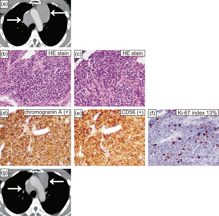
FIGURE 2.

Computed tomography (CT) taken for the current case again showed a mass in the anterior mediastinum (a). Microscopic findings of this case's needle biopsy specimen (hematoxylin–eosin staining and immunohistochemical findings, (b)–(f)). Hematoxylin–eosin staining findings comprised relatively homogeneous cells with weak atypia (b), (c). Hematoxylin–eosin staining showed one mitotic count at 2 mm2 (b) and no necrosis (c). Immunohistochemical staining was positive for chromogranin A (d) and CD56 (e). The Ki‐67 index was 13% (f). CT demonstrating the best response to first‐line chemotherapy showing shrinkage within the stable disease range (g). The magnification was 400× (b)–(f). HE, hematoxylin and eosin.
