
Figure 4.
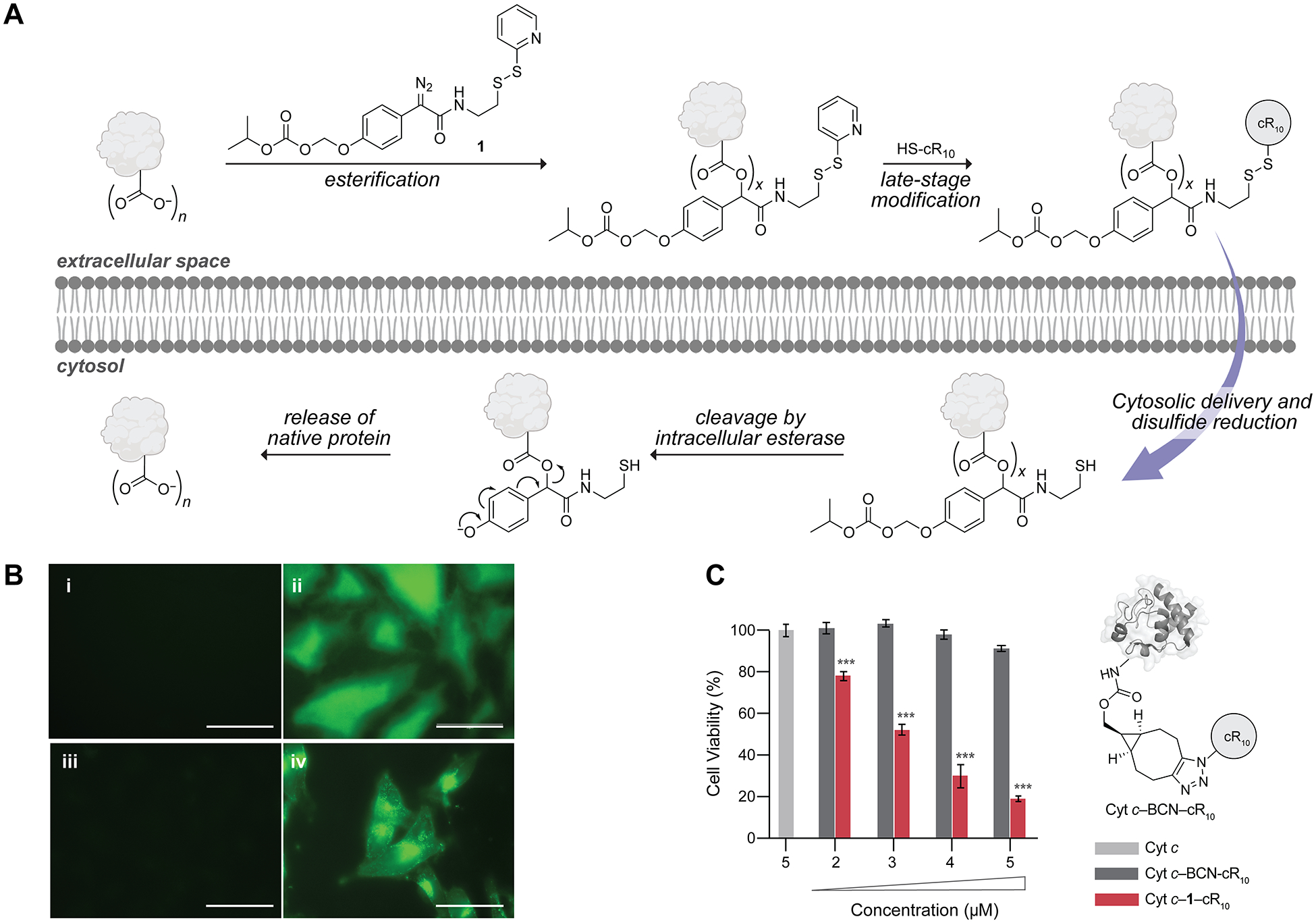
Cellular delivery of proteins. (A) Putative mechanism of cytosolic protein delivery via bioreversible modification with cR10. n refers to the total number of carboxyl groups; x refers to the number of ester labels. (B) Epifluorescence images of 5-μM (i) GFP, (ii) GFP–1–cR10, (iii) Cyt c–F, and (iv) Cyt c–F–1–cR10 incubated with live HeLa cells for 1.5 h (i, ii) or M21 cells for 2.5 h (iii, iv) in the presence of FBS-supplemented DMEM. λex = 488 nm and λem = 500 nm. Scale bars: 50 μm. (C) Viability of M21 cells upon a 41-h treatment with Cyt c (light gray), Cyt c–BCN–cR10 (dark gray, schematic view is shown), or Cyt c–1–cR10 (red). Values are the mean ± SD; ***p ≤ 0.001. For experimental details, see Figures S43, S44, and S47–S49.