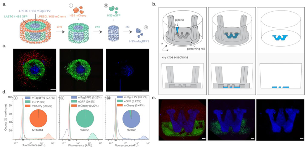
Figure 4 –

Spatiotemporally controlled liberation of single-cell suspensions of HS5 human stromal cells from complex trilayered materials through multiplexed sortase-based degradation. (A) Three distinctly fluorescent cell types were released from different sortase-degradable hydrogel fractions, with release fidelity was quantified by flow cytometry. (B) Arbitrary hydrogel geometries were constructed using open-microfluidic gel patterning through well-plate inserts. (C) Maximum intensity projections (MIPs) of 10x confocal images showing sortase-degradable bullseyes prior to degradation (left), following 4S9 treatment (center), and after 2A9 treatment (right). (D) Flow cytometry quantification of liberated cells following sequential treatment of 4S9 (left), 2A9 (center), and 5M (right) reveals that highly specific cell capture can be obtained from three different gel subregions with minimal off-target release. (E) Degradation of an arbitrary pattern as illustrated by MIPs of hydrogels patterned in the University of Washington Logo prior to degradation (left), following 4S9 treatment (center), and 2A9 treatment (right). Scale bars = 1 mm.
