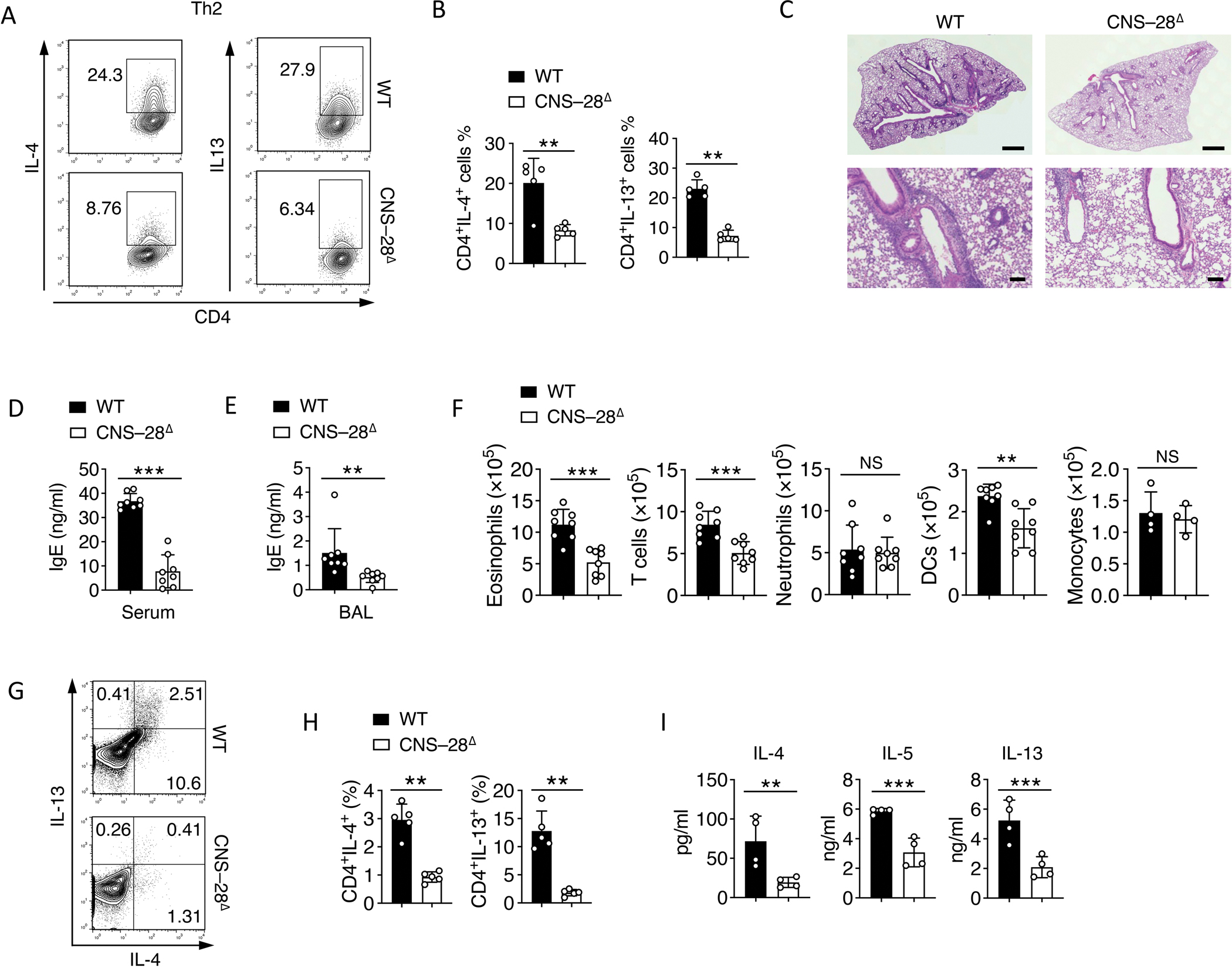
Fig 6. Reduced type 2 responses by deletion of CNS–28.

(A-B) Naïve CD4+ T cells from WT and CNS–28Δ mice were stimulated under Th2 condition and harvested at 72 hours. Intracellular staining of indicated cytokines was measured by (A) flow cytometry and (B) Quantification.
(C) Hematoxylin-and-eosin staining of lung-tissue sections of WT and CNS–28Δ mice, assessed after 10 days of HDM challenge. Scale bar, top row, 0.5 mm; bottom row, 100 μm.
(D-E) ELISA of IgE in the (D) serum and (E) bronchoalveolar lavage (BAL) fluid of WT and CNS–28Δ mice 10 days after HDM challenge.
(F) Frequency of inflammatory cells in the lung tissue of WT and CNS–28Δ mice, assessed at 10 days after HDM challenge.
(G) Flow cytometry analysis and (H) Quantification of type 2 cytokines in CD4+ T cells isolated from the lung 10 days after HDM challenge.
(I) Recovered cells from the lung 10 days after HDM challenge were restimulated by HDM. The levels of type 2 cytokines levels in medium were assessed by ELISA after 3 days of restimulation.
Data are representative of at least two independent experiments (A-C, G-I) or pooled from two independent experiments (D-F). **p < 0.01, ***p < 0.001, NS: not statistically significant. (Student’s t test, error bars represent SD).
