Fig. 8.
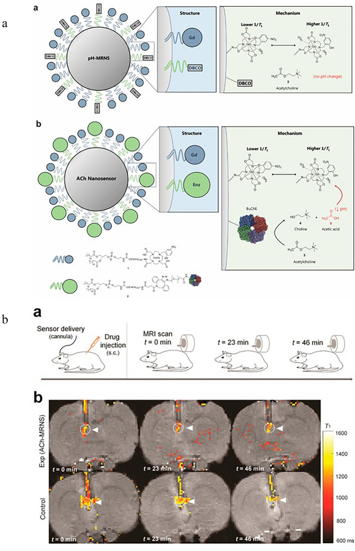
a Schematic illustration of the structure and mechanism of constructed nanosensor. a pH-MRNS. pH-sensitive contrast agents were conjugated to the DSPE-PEG [1, 2-distearoyl-sn-glycero-3-phosphoethanolamine-Poly(ethylene glycol)] lipids and coated on the surface of the lipophilic core [ACh is not hydrolyzed to alter local pH without coimmobilized BuChE (butyrylcholinesterase)]. b ACh-MRNS. pH-sensitive contrast agents and BuChE were covalently conjugated to the DSPE-PEG lipids and coated on the surface of the lipophilic core (the BuChE catalyzes the hydrolysis of ACh to Ch and acetic acid, resulting in a drop in local pH, which triggers a conformational switch of the contrast agent—one more water molecule coordinated to one Gd(III) chelate in acidic conditions, which leads to an increased contrast agent relaxation rate). Reprinted with permission from Ref. [129]. Copyright (2018) American Chemical Society. b ACh detection in vivo. a—Experimental procedure—subcutaneous administration of drug and nanosensor delivery through the cannula and three MR scans at different times. b—Exemplary coronal brain slices presenting ACh detection at different times. Reprinted with permission from Ref. [129]. Copyright (2018) American Chemical Society

