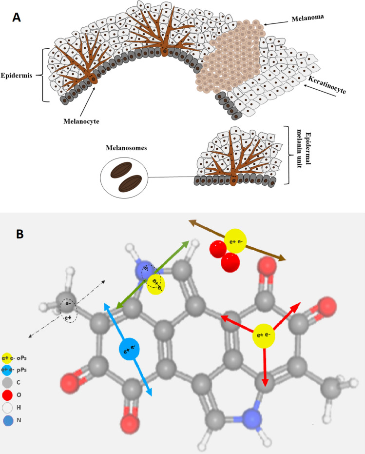
Figure 1.
Schematic view of melanocytes and the formation of a melanoma lesion in the epidermal layer. (A) Normal melanocytes have a regular dendritic shape and form many branching processes extending between numerous keratinocytes (left). One melanocyte reaches between 36 and 40 keratinocytes, forming an epidermal melanin unit (EMU), with a balanced proportion of one melanocyte to each of 8–10 keratinocytes in the basal layer of the epidermis. In a melanoma lesion (right), melanocytes lose their dendricity and become malignant and amoeboid in shape, changing their cell–cell contacts by expressing different adhesion molecules (N-cadherin instead of E-cadherin expressed by melanocytes)7. Melanosomes store the melanin granules produced by melanocytes, which can be distributed among surrounding keratinocytes to protect them from UV radiation damage. (B) Pictorial illustrations of positron annihilations in the melanin molecule. Carbon, oxygen, hydrogen, nitrogen, p-Ps and o-Ps atoms are indicated in colours explained in the legend. Dashed arrows indicate photons from direct electron–positron annihilation. Red arrows indicate photons from o-Ps atom self-annihilation. Blue arrows show photons from p-Ps self-annihilation. Green and brown arrows indicate o-Ps decay via the pick-off and conversion processes, respectively.

