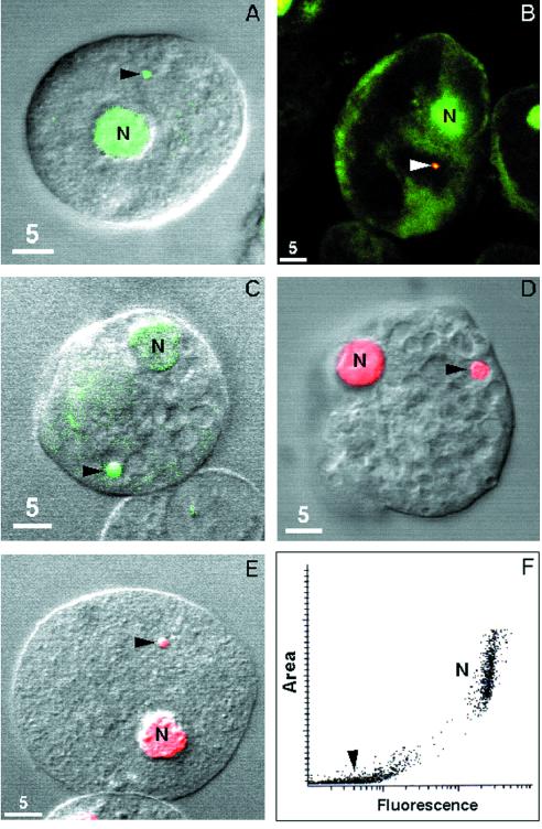
FIG. 1.
Confocal micrographs of nuclei (N) and cryptons (arrows) of E. histolytica trophozoites stained with DNA-binding fluorochromes, including sytox green (green [A and B]), acridine orange (green [C]), and propidium iodide (red [E]) (27). The crypton was also stained with anti-Hsp60 antibodies (yellow [B]) and a mouse monoclonal antibody to double-stranded DNA (red [D]) (13, 17). (F) Scatter diagram shows area and propidium iodide staining of the crypton and nuclei (14).

