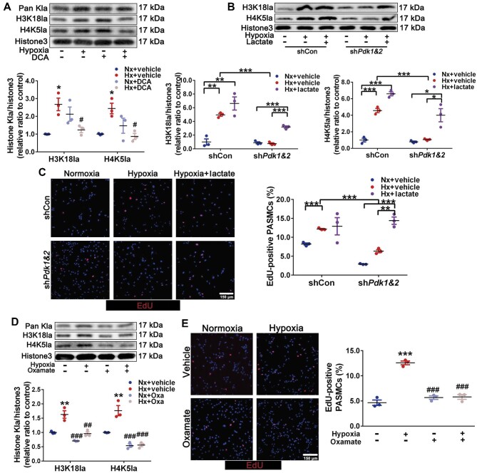
Figure 5.
Histone lactylation induced by lactate accumulation promotes PASMC proliferation. (A) Immunoblots for Pan Kla, H3K18la, H4K5la, and histone H3 in PASMCs exposed to normoxia and hypoxia with or without DCA (10 mM) incubation. (B and C) PASMCs with shCon or shPdk1&2 were exposed to normoxia, hypoxia, and hypoxia + lactate (10 mM). (B) Immunoblots for H3K18la, H4K5la, and histone H3. (C) Representative EdU (red) images, with quantification of the percentage of EdU-positive nuclei. Scale bar, 150 μm. Data represent mean ± SEM; *P < 0.05, **P < 0.01, ***P < 0.001. (D and E) PASMCs were exposed to normoxia and hypoxia with or without oxamate (50 μM) incubation. (D) Immunoblots for H3K18la, H4K5la, and histone H3. (E) Representative EdU (red) images, with quantification of the percentage of EdU-positive nuclei. Scale bar, 150 μm. Data represent mean ± SEM; *P < 0.05, **P < 0.01, ***P < 0.001 vs. Nx; #P < 0.05, ##P < 0.01, ###P < 0.001 vs. Hx; two-way ANOVA and Bonferroni's post-test. Oxa, oxamate; histone3, histone H3.

