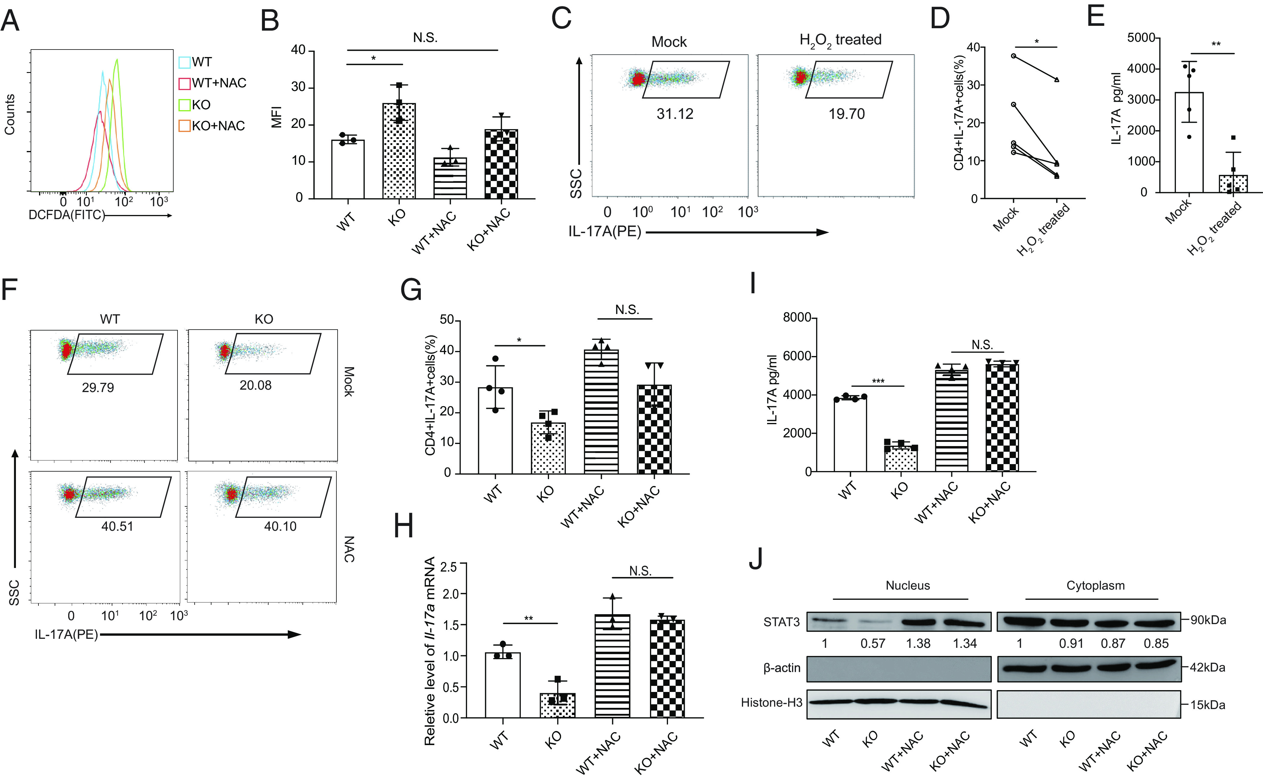
Fig. 3.

PRAK-regulated reactive oxygen species (ROS) elimination contributes to Th17 cell differentiation. Naïve CD4+T cells from WT and KO mice were cultured under Th17 polarizing conditions with or without the addition of 5mM N-acetyl-L-cysteine (NAC) or 4nM H2O2. All assays were performed at day 3. (A and B) Measurement of cellular ROS in WT and KO Th17 cells using the fluorescent probe DCFDA / H2DCFDA. Representative histogram (A) and mean fluorescence intensity (MFI) (B) from three independent experiments are shown. (C–E) Inhibition of WT Th17 cell differentiation by H2O2. Representative dot plots of intracellular IL-17A staining (C). Frequencies of IL-17A+ cells in the culture (n = 5) (D). IL-17A concentration in the supernatant as determined by ELISA (n = 5) (E). (F–J) Rescue of Th17 cell differentiation defect of Prak-deficient T cells by NAC. Representative dot plots of intracellular IL-17A staining (F). Frequencies of IL-17A+ cells the various cultures (n = 4) (G). Relative levels of Il-17a mRNA assessed by quantitative RT-PCR (n = 3) (H). IL-17A concentration in the culture supernatant (n = 4) (I). STAT3 in nuclear and cytoplasmic fractions of Th17 cells induced under various conditions. Representative blots from three independent experiments are shown (J). The numbers indicate the relative levels of expression normalized against histone H3 or β-actin. The statistics was performed using Student’s t test. ∗P < 0.05; ∗∗P < 0.01; ∗∗∗P < 0.001; N.S. not significant.
