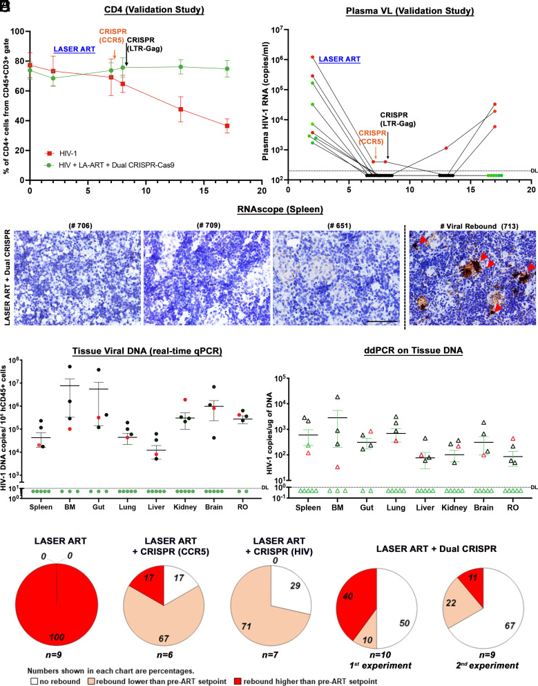
Fig. 4.
Viral and Immune profiles in HIV-1-infected and treated humanized mice of the validation study. (A) Flow cytometric evaluations of human CD4+ T cells in hu-mice assayed before (0) and 2, 7, 8, 13, and 17 wk postinfection from CD45+CD3+-gated populations. ART-treated and dual CCR5-HIV-1 CRISPR-administered mice showed restoration of absolute numbers of CD4+ T cells at the study end. (B) Plasma HIV-1 RNA copies of Hu-mice treated with ART and two sequential treatments of CRISPR-Cas9-targeting CCR5 and LTR-Gag indicated that six out of nine mice had no evidence of viral rebound in the plasma at 17 wk (study end). The sensitivity of detection was at 140 copies/mL after dilution factor adjustment. (C) Representative results from RNAscope assay revealed that 5 LASER ART and dual CRISPR-treated animals (numbers 706, 709, 622, 651, and 674) failed to demonstrate viral RNA amplification. The right panel shows one mouse spleen with viral rebound (#713). Images were captured at 40× magnification. (D) HIV-1 DNA analyses in the gag region using ultrasensitive semi-nested real-time qPCR assays from all the tissues of dual-treated animals (n = 9). Same five/nine animals shown in green closed circles showed no amplification of virus from all the tissues analyzed. The detection limit of the assay is 10 copies. One mouse (# 712) which was undetectable in plasma viremia was found to be HIV+ in tissue PCRs (shown in red closed circle). We could not find enough tissues from some mouse gut and BM for PCR, and the samples are missing from the datasets. The other three rebound mice (numbers 705, 707, and 713 shown in black circles) were found to be highly positive. The data represent mean ± SEM for each group. (E) ddPCR analysis of viral DNA from the nine dual-treated mice. The same five animals showed complete viral elimination in each of the tested tissues. One mouse (#712) which was undetectable in plasma viremia was found to be HIV-1 positive in tissue PCRs as shown in red open triangle. These results provide evidence of complete viral elimination in five mice (#s 706, 709, 622, 651, 674, green open triangle) and had no viral DNA detected in any tissues analyzed. (F) Plasma HIV-1 RNA copies of individual animals at 2WPI were compared to plasma HIV-1 RNA copies of the same animals at the study end, at 17WPI. The numbers of animals showing a lack of viral rebound (white), having rebound viral loads lower (salmon) or higher (red) than pre-ART setpoint are shown as a percentage for each of the ART-treated group. The numbers of animals in each category are indicated (n = x). To validate our findings in the ART and dual CRISPR treatment group (first evaluation) we conducted another independent experiment with a new set of nine humanized mice (second evaluation). Taken together, a total of 11 out of 19, ART and dual CRISPR-treated animals showed undetectable viral RNA at 17WPI (58%).

