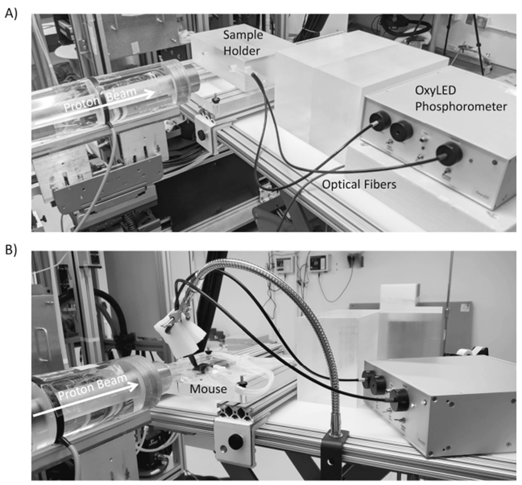
FIG. 1.

Panel A: Proton irradiation setup. A photo of the measurement setup used for ultrafast measurement of oxygen partial pressure in solutions during proton irradiation. Protons enter from the left side of the image through the beam collimator. The phosphorometer used to monitor oxygen partial pressure in either glass vials or in mice, is shown on the right. The sample holder for the vials was an acrylic block designed to hold the glass vial in the beam and position the optical fibers orthogonal to the beam axis to provide excitation and collect emission from the PtG4 phosphorescent probe in solution. Panel B: For in vivo experiments, the sample holder block was replaced by a heating pad to which the mice were taped while breathing anesthetic gas.
