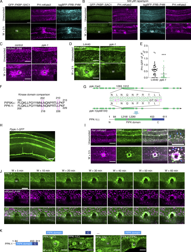
Figure S2.
PtdIns(4,5)P2 generation depends on PtdIns4P, PI4K, and PPK-1 activity, related to Fig. 2. (A) Representative single-plane confocal images showing GFP::FKBP::SAC-1, PH::mKate2, and tagBFP::FRB::P4M without rapamycin treatment before and after wounding. Pcol-19-PH::mKate2(zjuSi321) II; Pcol-19-P4M::FKBP12(mTOR)::tagBFP; Pcol-19-GFP::FKBP1A(FKBP 2-108AA)::SAC1(2-517AA)(zjuEx1859) transgenic animals were used for wounding and imaging. “W” indicates the wound site. Scale bar: 10 µm. (B) Representative single-plane confocal images showing GFP::FKBP::SAC-1, and PH::mKate2 with FRB, as well as tagBFP::FRB::P4M and PH::mKate2 without FKBP before and after wounding. Worms are treated with rapamycin 100 µM). Pcol-19-PH::mKate2(zjuSi321) II; Pcol-19-GFP::FKBP1A(FKBP 2-108AA)::SAC1(2-517AA)(zjuEx1865) and Pcol-19-PH::mKate2(zjuSi321); Pcol-19-GFP::FKBP1A(FKBP 2-108AA)::SAC1(2-517AA)(zjuEx1865) transgenic animals were used for wounding and imaging. Scale bar: 10 µm. (C) Representative confocal images of mKate2::P4M before and after wounding with ppk-1 RNAi treatment. Pcol-19-mKate2::P4M(zjuSi333) animals were used for wounding and imaging. Scale bar: 10 µm. (D) Representative confocal images of PH::GFP before and after wounding with ppk-1 RNAi treatment. Pcol-19-PH::GFP(zjuSi175) transgenic animals were used for wounding and imaging. Scale bar: 10 µm. (E) Quantitation analysis of the PH::GFP intensity ratio (ΔFw/Fuw) at 3 h after wounding (D). The error bars represent the mean value ± SD (n = 41 and 33), one-way ANOVA multiple comparisons test, ***P < 0.001. (F) Comparison of the amino acid sequence of human PIP5K1A and C. elegans PPK-1. (G) Experimental design of ppk-1(syb6134) double point mutation based on the comparison of the nucleotide sequence of ppk-1(wt) and ppk-1(syb6134). Below is the secondary structure of ppk(syb6134) mutation. (H) Representative confocal images of Pppk-1::GFP showing the expression of ppk-1 in multiple tissues of young adult C. elegans. (I) Representative single-plane confocal images of myr::mKate2 and GFP::PPK-1 colocalization before and after wounding. Pcol-19-myr::mKate2(zjuSi46); Pcol-19-GFP::PPK-1(zjuSi367) transgenic animals were used for wounding and imaging. White dotted boxes indicate the zoom-in region. Magenta arrows indicate the colocalized puncta. Scale bar: 10 µm. (J) Representative time-lapse confocal images of the recruitment of GFP::PPK-1 and mKate2::P4M between 5 to 60 min after wounding. Pcol-19-GFP::PPK-1(zjuSi367); Pcol-19-mKate2::P4M(zjuSi333) transgenic animals were used for wounding and imaging. Scale bar: 10 µm. (K) Diagram of PPK-1 secondary structure and representative confocal images of the recruitment of truncated PPK-1 to the wound site. Pcol-19-PPK-1(1–453)::GFP(zjuEx2147), Pcol-19-GFP::PPK-1(c)(zjuEx2142), Pcol-19-PPK-1(1–85)::GFP(zjuEx2265,) Pcol-19-PPK-1(84–457)::GFP(zjuEx2267) transgenic animals were used for wounding and imaging. Scale bar: 10 µm.

