
Figure 8.
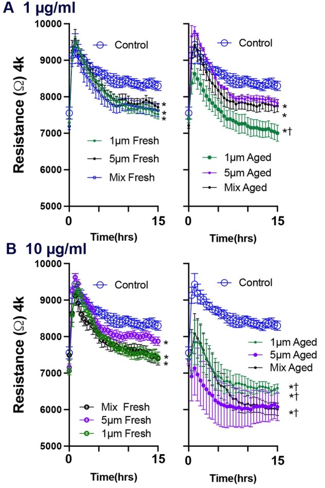
UV-aging enhances loss of barrier integrity in A549 cell treated with polystyrene microspheres. After achieving a stable resistance baseline plateau, cells were treated with the different concentrations/sizes of microspheres. Concentrations of 1 (A) and 10 μg/ml (B) were used. The barrier function responses in confluent A549 cells treated with polystyrene microspheres are showed in each figure for the first 15 h following cells treatment. Each figure compares 1 µm, 5 µm, and a mix of both sizes. Photoaged microspheres, the figures to the right of the fresh particles, were invariably more potent for both barrier and wound healing responses. N = 7 per group. Three-way ANOVAs were conducted to consider the effects of photoaging, size, and time. Asterisks (*) indicate a significant effect of microspheres compared with control and cross (†) indicated significant effect between photoaged and pristine microspheres.