Fig. 2.

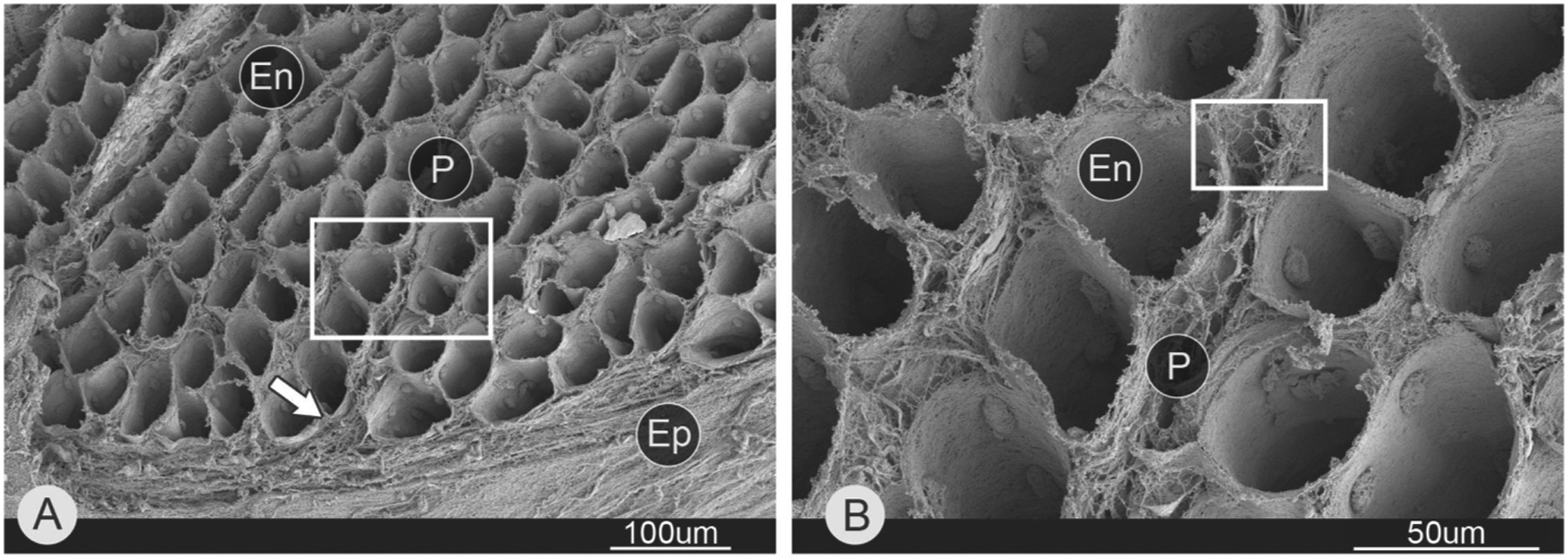
Scanning electron micrograph of endomysial connective tissue within skeletal muscle. This image was generated by scanning electron microscopy of a muscle whose fibers were removed by acid digestion. (A) Mouse lateral gastrocnemius. While boxed area shown in B. Arrow points to epimysium. (B) Magnified area from A. Abbreviations: endomysium (En), perimysium (P), and epimysium (Ep). (Micrograph reproduced from reference Sleboda et al., 2020).
