
Fig. 5.
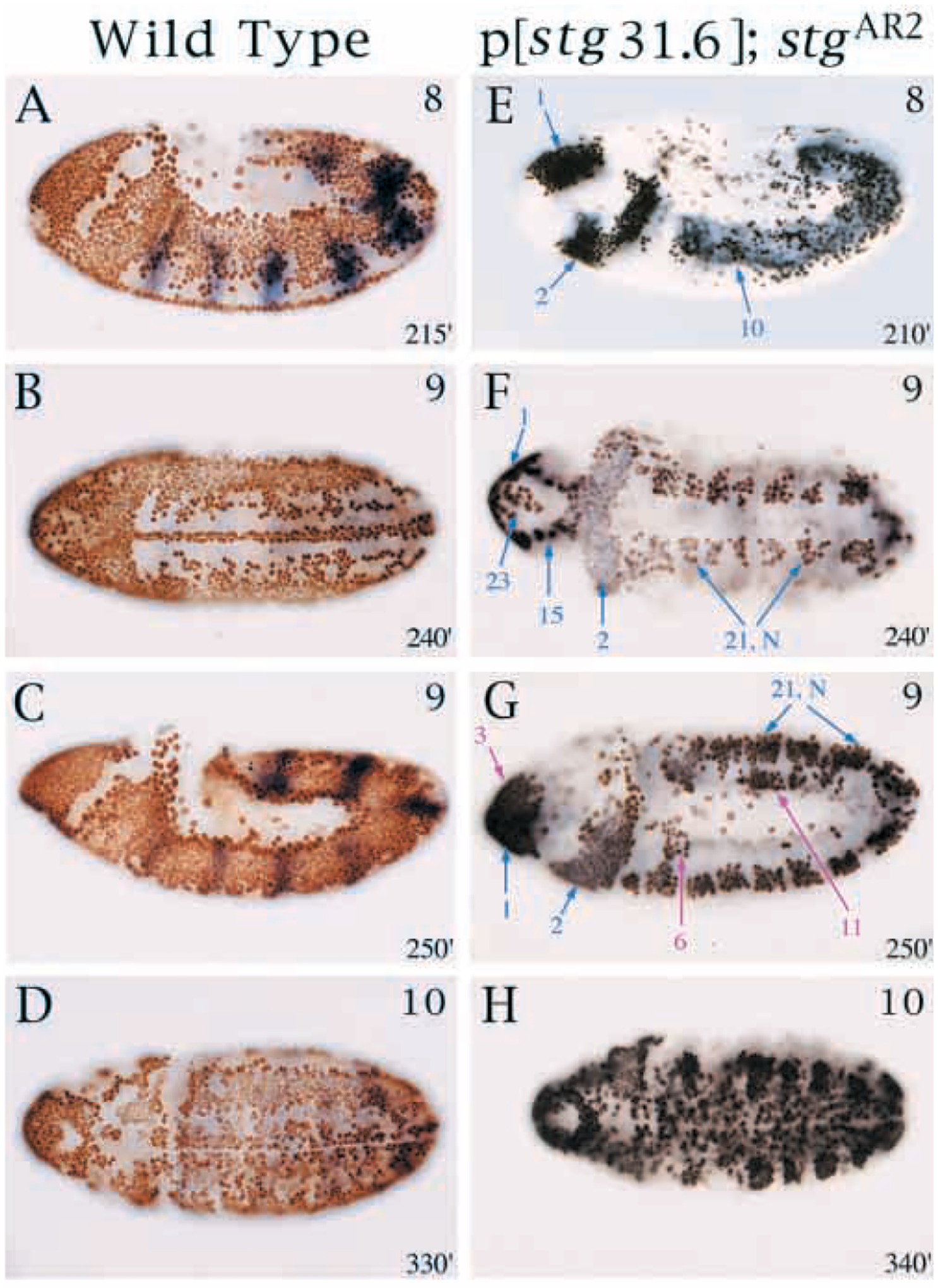
Embryonic cell cycles driven by a 31.6 kb string transgene. (A-D) Normal BrdU incorporation in control P[w+STG31.6]; stgAR2/TM3 Sb (P[ry+ ftz -lacZ]) embryos. (E-H) BrdU incorporation driven by the 31.6 kb transgene in a homozygous string null background; P[w+STG31.6]; stgAR2. All embryos are labeled with BrdU (brown or black stain) for 1 hour at 25°C and anti-β-gal antibody (blue) to detect the balancer. Approximate stages are indicated in upper right corner and age in minutes AED at 25°C is indicated in bottom right corners. Note views of embryos: (A,C,E,G) lateral; (B,D,F,H) ventral. (E-G) Mitotic domains driven by the 31.6 kb transgene that are also activated by individual PSEs are indicated by blue arrows. Mitotic domains in which cell division is driven by the 31.6 but not by the individual lacZ lines are indicated by fuchsia arrows and include cycle 15 MD, 3, 6 and parts of 11. BrdU incorporation in cycle 14 MDs 11 and 14, and cycle 15 MD 19 occurs but is inconsistent (data not shown). (H) Additional cycle 15 domains incorporate BrdU.