Figure 3.

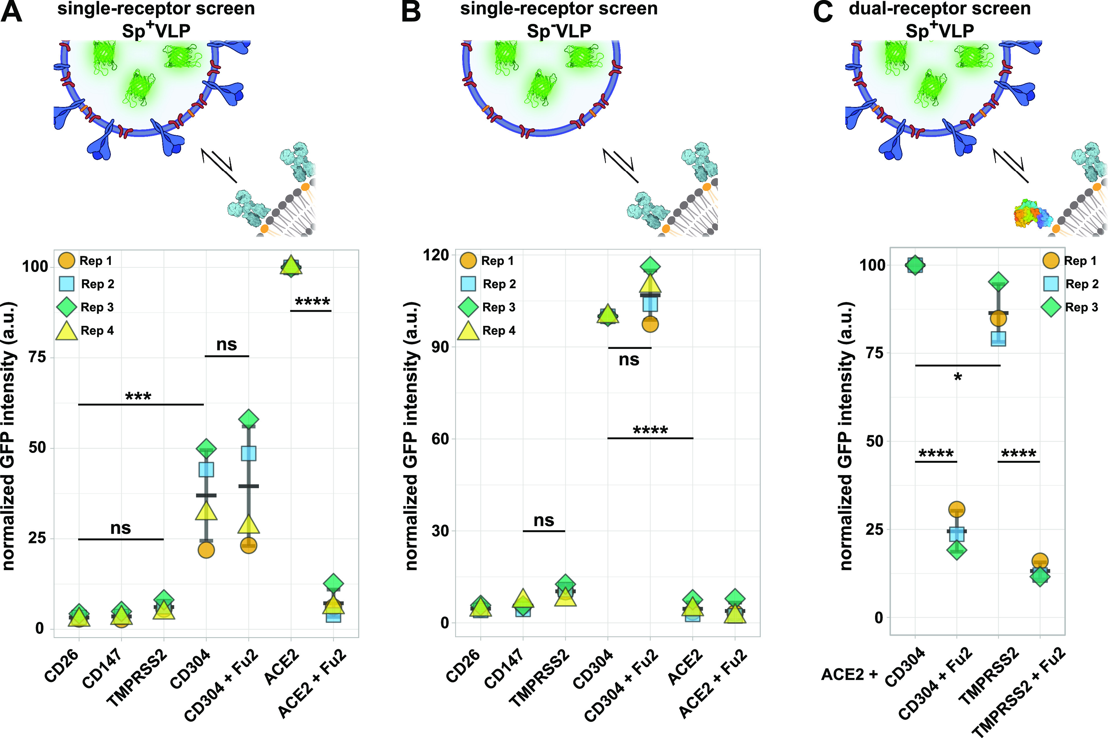
Receptor screening using fBSLBs. (A) Application of fBSLBs to study interaction between VLPs and different host-binding partners using flow cytometry. Scheme depicting fBSLBs with a single protein interacting with Sp+VLPs. Plot shows the ACE2-normalized medians of four different biological replicates (marked by different colors and shapes) from populations with ≈10 000 data points. The error bars show the standard deviation. Besides ACE2 (mean, 100), specific but less pronounced binding was also observed for CD304 (mean, 37), even when Sp+VLPs were blocked with Fu2 nanobody (mean, 40). (B) Scheme depicting fBSLBs with single protein interacting with Sp–VLPs. Plot shows the CD304-normalized medians of four different biological replicates (marked by different colors and shapes) from populations with ≈10 000 data points. Despite the absence of Spike, strong binding to CD304-fBSLBs was observed, even when Sp–VLPs were pretreated with Fu2 nanobody. (C) Dual receptor screen using fBSLBs and flow cytometry. Scheme depicting fBSLB coated with two proteins interacting with Sp+VLPs. BSLBs were functionalized simultaneously with equimolar concentrations of ACE2 and CD304 or TMPRSS2. Plot shows the ACE2+CD304-normalized medians of three different biological replicates (marked by different colors and shapes) from populations with ≈7000 data points. Despite blocking the interaction between Sp+VLPs and ACE2 with Fu2, there was still residual binding due to CD304 (mean, 24).
