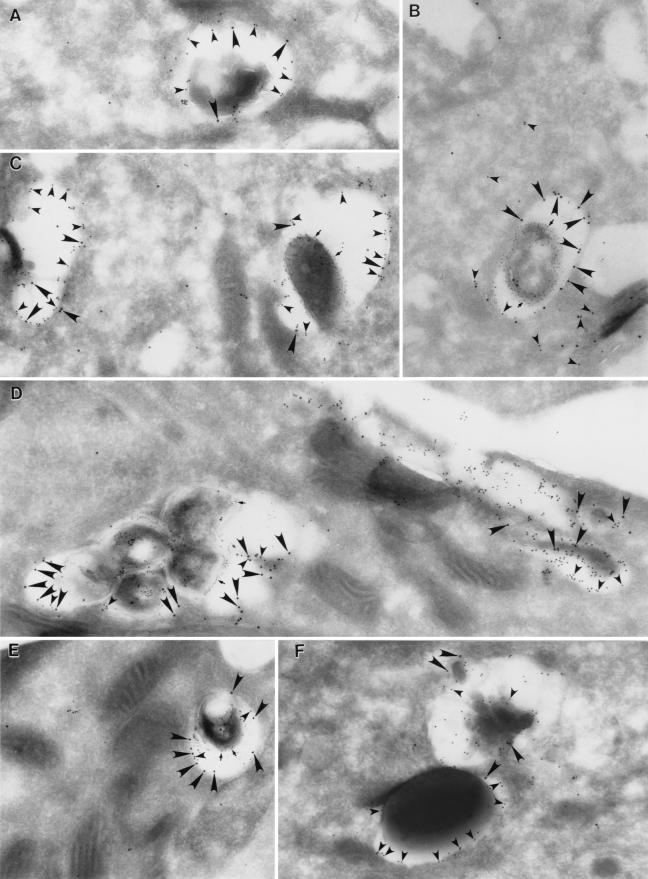
FIG. 10.
In HeLa cells expressing Rab7 Q67L, Rab7 Q67L colocalizes with LAMP-1 on cytoplasmic vesicles, but LAMP-1 remains absent from L. pneumophila and M. tuberculosis phagosomes. HeLa-Rab7 Q67L cells were fixed 2 h after infection with wild-type (A and B) or avirulent L. pneumophila (C), as described in the legend of Fig. 8, or 2 days after coinfection with live M. tuberculosis and latex beads (D, E, and F), as described in the legend of Fig. 9, and processed for immunoelectron microscopy. (A) Rab7 Q67L (15-nm immunogold particles; large arrowheads) colocalized extensively with LAMP-1 (10-nm immunogold particles; small arrowheads) on vesicles that appeared to be autophagosomes or multivesicular bodies. The absence of LPS excludes the possibility that the vesicle shown contains L. pneumophila. (B) L. pneumophila phagosomes often stained richly for Rab7 Q67L (15-nm gold particles; large arrowheads) but showed little or no staining for LAMP-1. LAMP-1 (10-nm gold particles; small arrowheads) was present on adjacent cytoplasmic vesicles. L. pneumophila LPS was stained with 5-nm gold particles (small arrows). (C) Avirulent L. pneumophila phagosomes frequently stained positive for Rab7 Q67L (15-nm gold particles; large arrowheads) and consistently stained intensely for LAMP-1 (10-nm gold particles; small arrowheads). L. pneumophila LPS was stained with 5-nm gold particles (small arrows). (D and E) M. tuberculosis phagosomes, like L. pneumophila phagosomes, frequently stained richly for Rab7 Q67L (15-nm gold particles; large arrowheads) yet acquired only low levels of LAMP-1 (10-nm gold particles; small arrowheads). Mycobacterial LAM was stained with 5-nm immunogold particles (small arrows). An autophagosome shown on the right side of panel D stains richly for both Rab7 Q67L and LAMP-1. (F) Latex bead phagosomes in these cells, on the other hand, had low to moderate levels of staining for Rab7 Q67L (15-nm gold particles; large arrowheads), but stained intensely for LAMP-1 (10 nm gold particles; small arrowheads). The latex bead phagosome shown in this panel has one Rab7 Q67L immunogold particle and approximately 20 LAMP-1 immunogold particles. An adjacent vacuole with multiple internal membranes stains positive for both Rab7 Q67L and LAMP-1. Magnifications are (A) ×41,300; (B) ×29,400; (C, D, and E) ×32,200; and (F) ×32,900.

