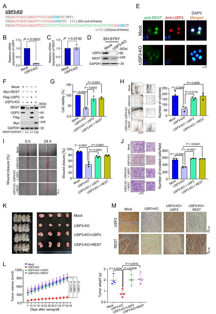
Fig. 7.
Loss of USP3 inhibits neuroblastoma tumorigenesis in vitro and in vivo. (A) Sanger sequencing data showing the disruption in USP3 gene sequence in SH-SY5Y cells (USP3-KO). (B) The effect of USP3 KO on the mRNA expression of USP3 and (C)REST was evaluated by qRT-PCR with specific primers. The relative mRNA expression levels are shown after normalization to GAPDH mRNA expression. Data are presented as the mean and standard deviation of four independent experiments (n = 4). A two tailed t-test was used, and P values are indicated. (D) Western blot analysis of the endogenous expression of USP3 and REST protein in USP3-KO was evaluated. GAPDH was used as the internal loading control. (E) Immunofluorescence staining of the endogenous expression of USP3 and REST in USP3-KO cell line. (F) Mock control, USP3-KO, and USP3-KO cells reconstituted with either USP3 or REST. Western blot analysis to validate the expression of USP3 and REST using USP3- and REST-specific antibodies. The protein band intensities were estimated using ImageJ software with reference to the GAPDH control band (REST/GAPDH) and presented below the blot. The cells from (F) were subjected to following experiments (G) cell viability by CCK-8 assay, (H) colony formation assay, (I) wound-healing assay and (J) transwell cell-invasion assay. Data are presented as the mean and standard deviation of three independent experiments (n = 3). One-way ANOVA followed by Tukey’s post hoc test was used, and P values are indicated. (K) Xenografts were generated by subcutaneously injecting mock control, USP3 KO, and USP3-KO cells reconstituted with USP3 or REST SH-SY5Y cells into the right flanks of NOD SCID gamma mice (n = 4/group). Tumor volumes were recorded. The right panel shows the tumors excised from the mice after the experiment. (L) The tumor volume was measured every 3 days for 43 days and is presented graphically. The tumor weight was recorded post-euthanization. Data are presented as the mean and standard deviation of four independent experiments (n = 4). Two-way ANOVA followed by Tukey’s post hoc test was used, and P values are indicated. (M) Xenograft tumors were embedded in paraffin and sectioned. The immunohistochemical analyses were performed with the indicated antibodies. Scale bar = 100 μm

