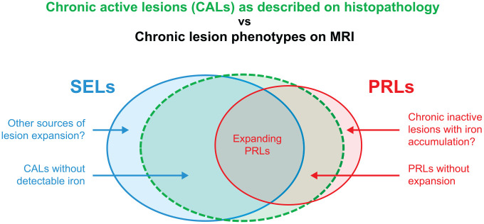
Figure 3.
Summary representation of CALs as described by histopathology versus imaging phenotypes of PRLs and SELs in MS.29,34
Figure is for representative purposes only, and the degree of overlap is not meant to convey a quantitative description of overlap between lesion types.
CAL: chronic active lesion; PRL: paramagnetic rim lesion; SEL: slowly expanding lesion.

