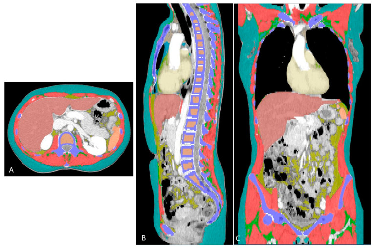
Figure 1.
Colored segmentations in the axial (A), sagittal (B) and coronal (C) views. The different colors show each compartment automatically segmented, as follows: red = skeletal muscle; green = intramuscular adipose tissue; cyan = subcutaneous adipose tissue; blue (aqua) = paracardial adipose tissue; green = epicardial adipose tissue; purple = bone; bronze = trabecular bone; brick red = liver; amber = spleen; and ivory = heart.

