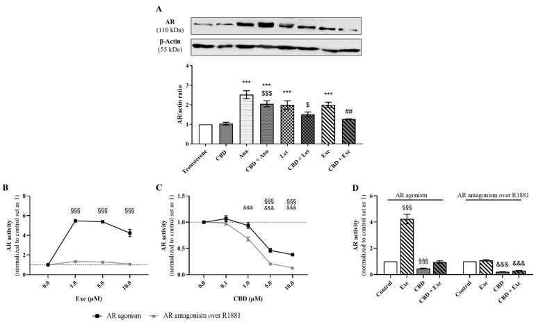
Figure 5.
Involvement of AR in the effects of the AIs, Ana, Let, and Exe, as well as of CBD and their combinations on breast cancer cells. (A) AR protein expression levels were evaluated by Western blot in MCF-7aro cells stimulated with T (1 nM) and treated with the AIs (10 µM), CBD (5 µM) or their combinations for 3 days. Cells treated only with T were used as control. A representative Western blot of AR and β-actin, as well as the densitometric analysis of AR expression levels after normalization with β-actin levels, used as loading control, are presented. (B–D) AR transactivation assay was performed in the AR-EcoScreen™ cells treated with Exe (0.1–10 µM; B), CBD (0.1–10 µM; (C) and their combination (D) in the presence or absence of R1881 (0.1 nM) over 24 h. Statistically significant differences between MCF-7aro cells treated with compounds and control (T) are represented as *** (p < 0.001), while differences between the combinations and each AI alone are indicated as ## (p < 0.01) and the differences between the combinations and CBD as $ (p < 0.05) and $$$ (p < 0.001). For transactivation assays, the statistically significant differences between the control and cells treated with compounds but without R1881 are expressed as §§§ (p < 0.001), while differences between the control and cells treated with compounds in the presence of R1881 are denoted by &&& (p < 0.001). The original Western blots are represented in Supplementary Figure S1C.

