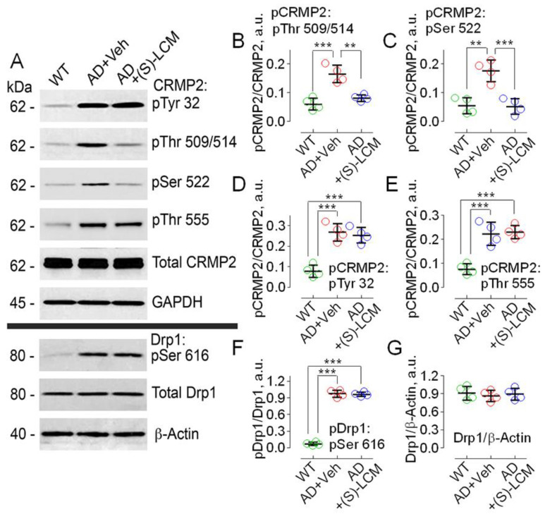
Figure 3.
Hyperphosphorylation of CRMP2 at Ser 522, Thr 509/514, Tyr 32, Thr 555, and hyperphosphorylation of Drp1 at Ser 616 in cultured cortical neurons from APP/PS1 mice compared to neurons from WT littermates. (S)-LCM (10 µM for 7 days) significantly reduced CRMP2 phosphorylation at Ser 522 and Thr 509/514, but not at Thr 555 and Tyr 32 or at Ser 616 of Drp1. Cortical neurons were isolated from P1 APP/PS1 (AD) and WT mice of both sexes and cultured for 12–14 days in vitro (12–14 DIV). In (A), representative immunoblots. (B–G), statistical summaries based on densitometry data. Where shown, neurons were exposed to 10 µM (S)-LCM for the last 7 days before the experiments. β-actin and GAPDH are loading controls. Here and in other experiments with (S)-LCM applied to cultured neurons, 0.01% DMSO was used as a vehicle (Veh). Green circles, neurons from wild-type (WT) mice; red circles, neurons from AD mice treated with a vehicle; blue circles, neurons from AD mice treated with (S)-LCM. Data are mean ± SD, ** p < 0.01, *** p < 0.001, N = 4 separate experiments with neurons from different platings. Data were analyzed by parametric one-way analysis of variance (ANOVA) with Holm–Sidak post-test.

