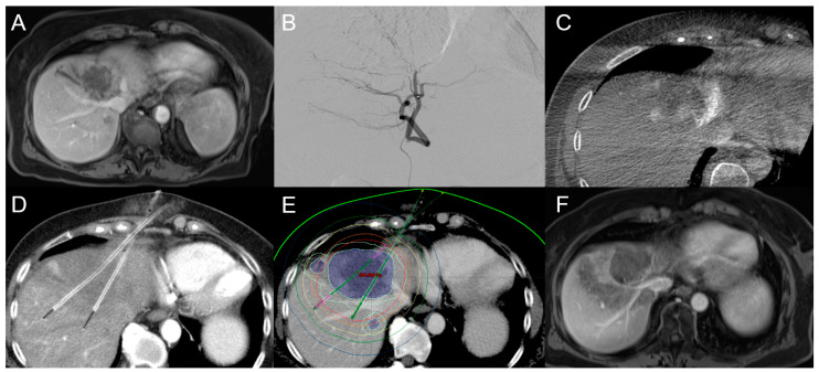
Figure 5.
An 84-year-old female patient with initial diagnosis of unresectable iCCA with a diameter of up to 6.5 cm in segment IVa close to the inferior caval vein confluence (A). (B) shows the embolization position before TACE and (C) shows the peri-interventional cone beam CT scan. After TACE, CT-HDRBT was performed (the colored lines represent the isodose boundaries) (D,E), while (F) shows the huge ablation zone covering most of the iCCA mass.

