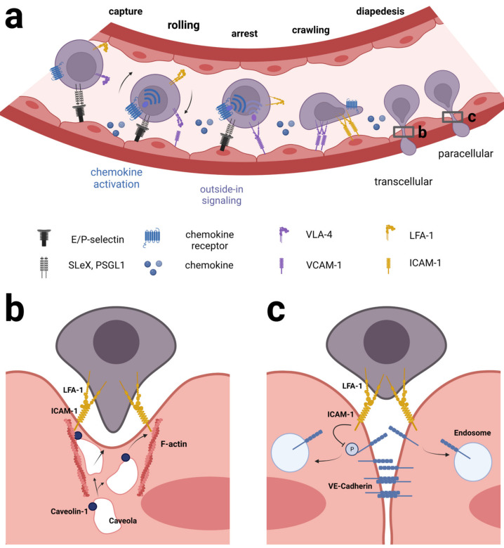
Figure 2.
Mechanism of leukocyte extravasation from blood vessels. (a) The process starts by capture/tethering, which mainly depends on endothelial E-and P-selectins binding sialyl-Le X ligands or P-selectin glycoprotein ligands on the leukocyte surface, leading to leukocyte rolling on the luminal surface of the endothelium. VLA-4 (α4β1) binding to VCAM-1 and LFA-1 (αLβ2) binding to ICAM-1 contribute to slowing down the rolling. A combination of chemokine activation and VCAM-1 and ICAM-1 binding increases the affinity of the integrins via inside-out and outside-in signaling, respectively. Additionally, the blood flow and the formation of catch bonds contribute to strengthening the LFA-1: ICAM-1 interaction. Eventually, this leads to arrest of the leukocyte. Following the formation of an LFA-1: ICAM-1 transmigratory cup, the leukocyte starts crawling and at the same time extends membrane protrusions to probe the endothelial surface to find a site permissive of transmigration. The last step of extravasation is diapedesis during which the leukocyte moves through the endothelium either through an individual EC cell (transcellularly) or through a space between neighboring EC (paracellularly) highlighted in (b,c), respectively. (b) transcellular diapedesis involves clusters of ICAM-1 on the endothelial cell, allegedly binding LFA-1 on the leukocyte. ICAM-1 is in physical contact with both F-actin filaments and the protein caveolin-1 on the surface of caveolae inside the endothelial cell. The caveolae gradually fuse to form a channel through which the leukocyte slides through. (c) In paracellular diapedesis, LFA-1 on the surface of the leukocyte binds ICAM-1 on two neighboring endothelial cells. ICAM-1 alters the phosphorylation status of key tyrosine residues in vascular-endothelial cadherin (VE-cadherin) causing VE-cadherin to be endocytosed by the endothelial cells. As a result, the adherens junctions between the endothelial cells are gradually unzipped as the leukocyte moves through. See text for further details. This figure was created with BioRender.com.

