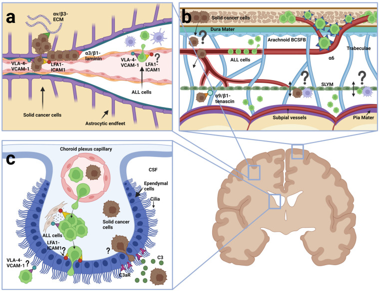
Figure 4.
Entry routes and integrins used by ALL cells (green) and metastatic cells from solid cancers (brown) (a) Postcapillary venule. Metastatic cells extravasate from venules in a manner dependent on VLA-4 (α4β1): VCAM-1 and LFA-1 (αLβ2)-ICAM-1. Once across, they either engage in vascular co-optive growth to which α3 and β1 integrins may contribute, or they establish metastasis in the parenchyma, which involves αVβ3. Although presumed to involve α4β1: VCAM-1 and αLβ2-ICAM-1, little is known of integrins involved in ALL extravasation from postcapillary venules. (b) section showing meningeal layers between the calvaria and the brain parenchyma. ALL cells use α6 integrin: laminin binding to migrate on the basement membrane of emissary vessels connecting calvarial bone marrow and the meninges. It is unknown if and how metastatic cells from solid cancers enter the meninges from calvarial bone marrow. Both ALL cells and metastatic cells are found in the dura and leptomeninges. Different integrins may be involved in the binding to meningeal components. It is unknown whether cancer cells of both types traverse or otherwise interact with the SLYM (indicated by a question mark). (c) choroid plexus (CP). ALL cells use α4β1: VCAM-1 and αLβ2: ICAM-1 to interact with stromal fibroblasts and studies show that they can cross the BCSFB. Metastatic cancer cells in the CSF use the complement component C3 binding the receptor C3aR on the ependymal cells to disrupt the BCSFB. This figure was created with BioRender.com.

