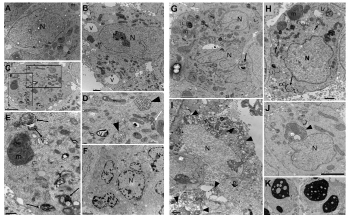
Figure 3.
Transmission electron microscopy of LNCaP clone FGC cells treated for 48 h with CC50 of fruit (B–F) and seed (G–K) Euterpe oleracea extracts. (A) Untreated cells. (B–E) Presence of large vacuoles (V), residual bodies (arrows); alteration in size and shape of mitochondria; swelling and loss of mitochondrial matrix (head arrows). (F) Cells with numerous vesicles (ve) and alteration of chromatin condensation in nuclei. (G–J) Presence of lipids (L), residual bodies (arrows), increase in the number of mitochondria with different shapes and sizes; mitochondria swelling, and loss of mitochondria cristae and matrix (head arrows). (K) Pyknotic nuclei. N: nucleus, m: mitochondrion. Scale bar is 0.5 µm in (D,E); 1 µm in (A,B,H,I,K); and 2 µm in (C,F,G,J).

