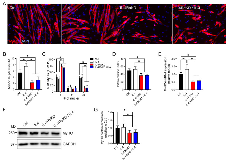
Figure 2.
IL-4Rα knockdown impaired myoblast fusion and differentiation in C2C12 cells. (A) Representative immunofluorescence images of MyHC (red) in the IL-4RαKD C2C12 myoblasts. Cell nuclei were counterstained with DAPI (blue). Myotube formation was inhibited in the IL-4RαKD C2C12 cells. In this experiment, C2C12 cells were transfected with control siRNA (Ctrl) or IL-4Rα siRNA (IL-4 RαKD) and grown in GM for 24 h. The medium was replaced by DM with or without recombinant IL-4 (10 ng/mL). After 72 h incubation, cells were fixed and stained. Scale bars: 50 μm. (B–D). In (B–D), the number of myonuclei per myotube; the percentage of myosin-positive cells containing 1, 2, or ≥3 nuclei; and the differentiation index were shown, respectively. These parameters of myoblast differentiation significantly decreased by IL-4Rα knockdown. N = 5 per group. (E–G) Reduction of IL-4/IL-4Rα signaling by IL-4Rα knockdown significantly suppressed the increased expression of MyHC. Cells were treated as described in (A). (E,G) show fold changes in mRNA and protein levels, respectively. N = 6 per group. Representative western blot analysis is shown in (F). N = 6 per group. Data represent mean ± SD. * p < 0.05.

