Figure 5.
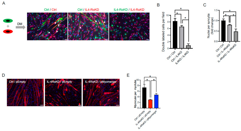
Supplementation of myomerger partially restored the impaired myoblast fusion capacity in IL-4RαKD C2C12 cells. (A–C) In a cell mixing assay, C2C12 cells were transfected with control (Ctrl) or IL-4Rα siRNA (IL-4RαKD) and labeled with green or red fluorescent lipid. Equal numbers of cells with each color were mixed and differentiated in DM for 72 h. (A) Representative images of cell mixing experiments. Arrowheads indicate double-labeled syncytia. Scale bars: 50 μm. (B,C) The number of double-labeled cells and the number of nuclei per syncytia were significantly increased in the Ctrl/IL-4RαKD group when compared to the IL-4RαKD/IL-4RαKD group. However, it did not reach the level of the Ctrl/Ctrl group. N = 5 per group. (D,E) One day after siRNA transfection, a myomerger expression vector (pMyomerger) or a control vector (pEmpty) was transfected. After 72 h of differentiation in DM, myoblast cells were immunostained or subjected to Western blotting analysis. (D) Representative immunofluorescence images of MyHC (red). Cell nuclei were counterstained with DAPI (blue). Scale bars: 50 μm. (E) The number of myonuclei per myotube in the IL-4RαKD/pMyomerger group was significantly greater than that in the IL-4RαKD/pEmpty group but still significantly less than that in the Ctrl/pEmpty group. N = 4 per group. Data are presented as mean ± SD. * p < 0.05.

