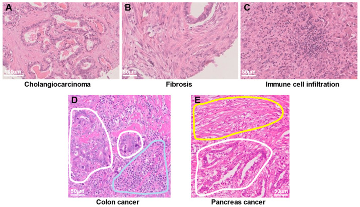
Figure 1.
Heterogeneous cell populations in human cancer tissue. (A–C) Hematoxylin and eosin (H&E) staining of the same cholangiocarcinoma tissue specimen reveals cancer cells (A), fibrotic tissue composed of fibroblasts (B), and infiltration of immune cells consisting mostly of lymphocytes (C). (D) H&E staining of colon cancer tissue shows cancer cells (areas surrounded by white lines) in close proximity to infiltrating immune cells (area surrounded by the blue line). (E) H&E staining of a pancreatic cancer specimen reveals fibrotic tissue (area surrounded by the yellow line) adjacent to cancer cells (area surrounded by the white line).

