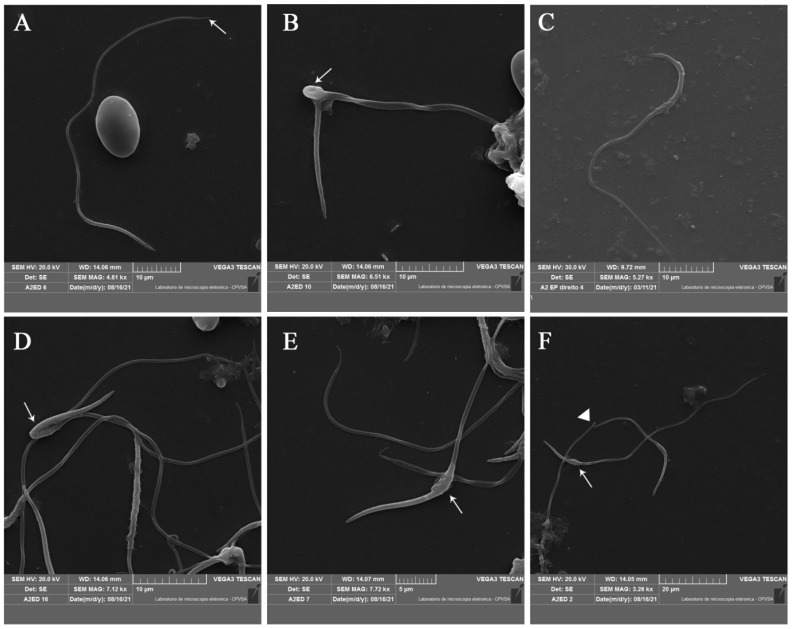
Figure 3.
Scanning electron microscopy of rhea (Rhea americana) spermatozoa from the epididymis. (A) Normal sperm with endpiece (white arrow). (B) Macrocephalic sperm, with two tails and bent midpiece (white arrow). (C) Sickle-shaped head. (D) Bent head. (E,F) Swelling at the base of the head (white arrows). (F) Broken tail (white triangle).

