
Fig. 4 |. Heat shock increases chromatin-binding fraction of HSF without affecting dwell time.
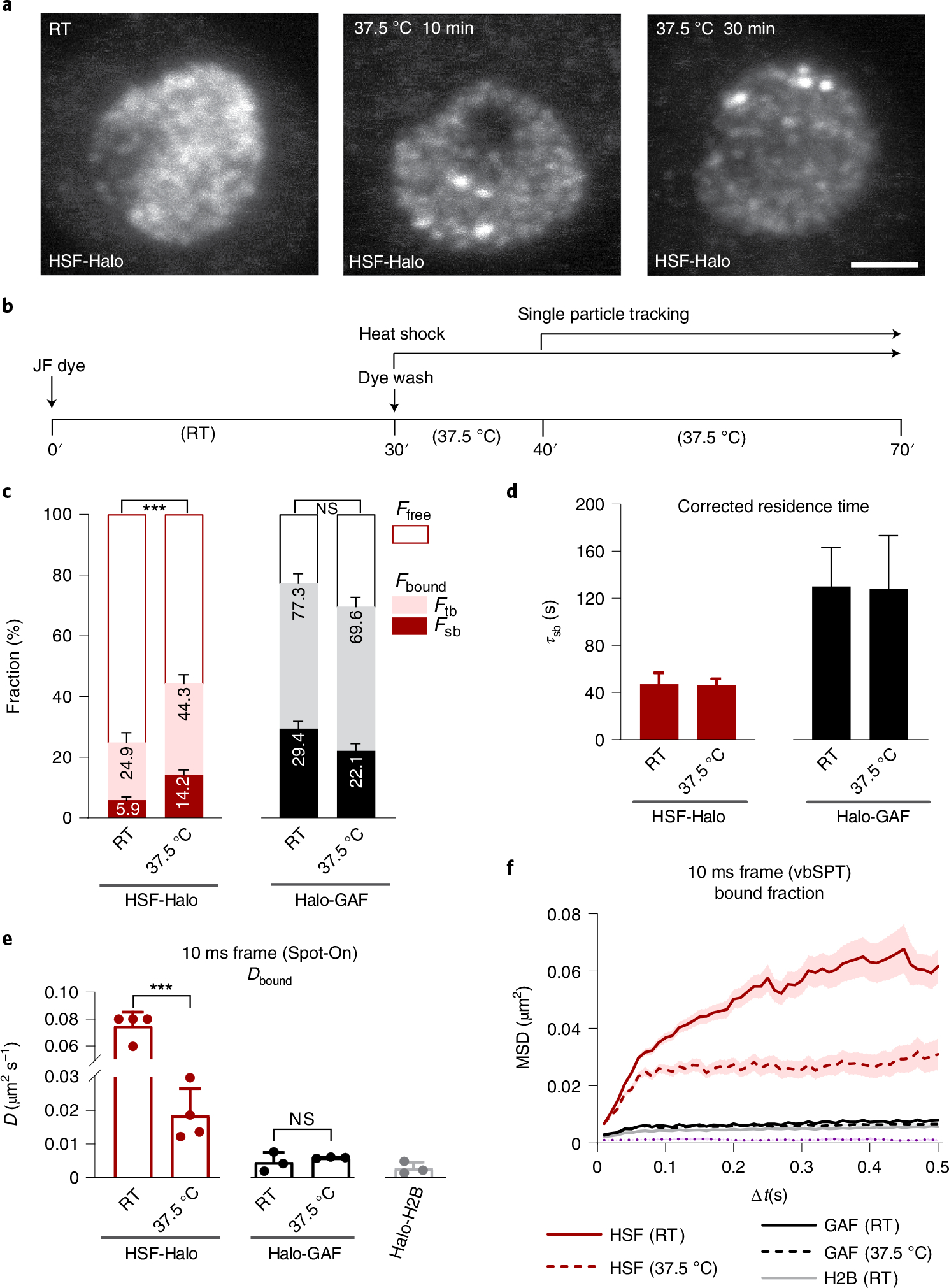
a, Maximum-intensity z-stack projection of HSF-Halo in fixed hemocytes. HSF-Halo forms several prominent foci on heat shock at 37.5 °C. Maximum projections of confocal z-stacks are shown. Scale bar, 2 μm. b, Flow chart of the heat shock and live-hemocyte imaging procedure. SPT starts 10 min after heat shock and continues over multiple cells (1–2 min per cell) for a total of 30 min with each sample. c, Global chromatin-bound fractions for HSF-Halo and Halo-GAF at RT and 37.5 °C, from fast tracking ( biological replicates for HSF-Halo, biological replicates for Halo-GAF) and slow tracking ( resamplings). Results are mean ± s.d. with error propagation for , two-sided, unpaired t-test for fast tracking. d, Corrected average residence times for HSF-Halo and Halo-GAF at RT and 37.5 °C. Error bars represent bootstrapped s.d. after resampling 100 times . e, Diffusion coefficients for bound fractions of HSF-Halo and Halo-GAF at RT and 37.5 °C, and Halo-H2B at RT derived using Spot-On. Results are mean ± s.d. ( biological replicates for HSF-Halo, biological replicates for Halo-GAF and Halo-H2B). , two-sided, unpaired t-test. f, Average MSD (mean ± s.e.) versus lag time of bound trajectories classified by vbSPT for HSF-Halo, Halo-GAF and Halo-H2B at RT and 37.5 °C and Halo-H2B at RT. See Extended Data Fig. 9a for a zoomed-in section for GAF and H2B.