Figure 5.
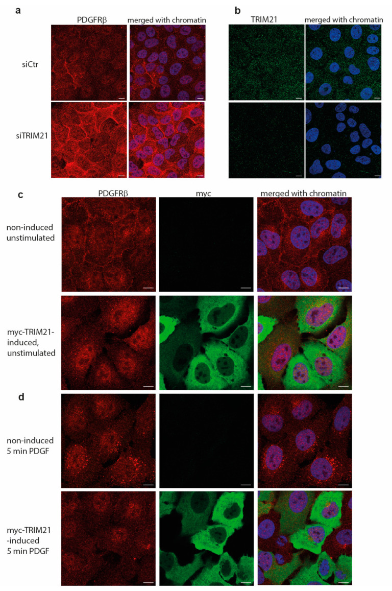
(a) Immunofluorescence staining of PDGFRβ in U2OS cells that were treated with control siRNA (“siCtr”) or depleted of TRIM21 protein (“siTRIM21”). Single channel staining of PDGFRβ is presented (red) and merged with nuclear staining with DAPI (blue). (b) Immunofluorescence staining of endogenous TRIM21 (green) shows weak cytoplasmic distribution in control cells (“siCtr”) and efficient knockdown in (siTRIM21) cells; staining merged with nuclei (blue) images are also presented. Confocal images in (a,b) were taken with 63× oil objective and zoom 0.5. (c,d) Immunofluorescence staining of PDGFRβ in myc-TRIM21-inducible U2OS cells cultured in 10% FBS after induction of TRIM21 before (c) or after (d) stimulation with PDGF-BB for 5 min. PDGFRβ staining is in the red; TRIM21 (myc) staining is in green; chromatin counterstained with DAPI is in the blue. Merged images for all three channels are presented on the right. Confocal images in (c,d) were taken with 63× oil objective and zoom 0.7. Scale bars are 10 µm.

