Figure 1.
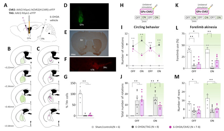
GPe photostimulation ameliorates circling behavior and forelimb akinesia in 6-OHDA-lesioned mice. (A) Schematic illustration of the experimental procedure for opsin expression in GPe and nigrostriatal DA lesion. (B) Illustration of the immunofluorescence spreading across the anteroposterior axis in the brain sections of 6-OHDA/ChR2 mice containing the GPe. (C) Placement of individual optical fiber across the anteroposterior axis of the GPe for all experimental groups. (D) Immunofluorescence image illustrating unilateral eYFP expression in GPe neurons. (E) DAB immunostaining for TH-positive fibers in the striatum of a lesioned mouse. (F) Immunostaining for TH+ cells in SNc of a lesioned mouse. (G) Percentage of TH+ cell loss in SNc in lesioned mice relative to Sham mice. (H) Photostimulation protocol used for the open field test. Testing was carried over 8 min with repeated sequences of light pulses stimulation (532 nm, 5 Hz, 5 ms, 3 mW) turned OFF and ON for 2 min. (I,J) Time course and a total number of spontaneous ipsilateral rotations during OFF and ON periods, respectively. (K) Photostimulation protocol was used for the cylinder test. Testing was carried over 6 min with repeated sequences of light pulses stimulation (532 nm, 5 Hz, 5 ms, 3 mW) turned ON and OFF for 1 min. (L) Percentage (%) of contralateral forelimb use during OFF and ON periods (number of contralateral forepaw contacts relative to the total number of forepaw contacts). (M) Total number of rears (forepaw contacts) during OFF and ON periods. Sham/controls, N = 6; 6-OHDA/TAG, N = 8; 6-OHDA/ChR2, N = 7–8. One mouse from the 6-OHDA/ChR2 group was excluded from the cylinder test (see Methods). Results are presented as mean ± SEM. * p < 0.05 and ** p < 0.01 significantly different from Sham mice. # p < 0.05 significantly different from 6-OHDA/TAG mice, Dunn’s test following a significant non-parametric Kruskal–Wallis test.

