Figure 3.
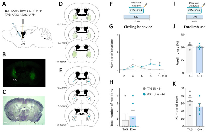
Unilateral GPe photoinhibition does not produce motor deficits. (A) Schematic illustration of the experimental procedure for opsin expression in GPe. (B) Immunofluorescence image illustrating bilateral viral expression in GPe neurons. (C) Cresyl violet staining showing bilateral fiber optic placement in the GPe. (D) Illustration of the immunofluorescence spreading across the anteroposterior axis in the brain sections of iC++ mice containing the GPe. (E) Placement of individual optical fiber across anteroposterior GPe axis for TAG and iC++ mice. (F) Photoinhibition protocol used in the open field test. Testing was carried out over 10 min with continuous light illumination (450 nm, 12 mW). (G,H) Time course and the total number of ipsilateral rotations over 10 min of testing, respectively. (I) Photoinhibition protocol used in the cylinder test. Testing was carried out over 6 min with continuous light illumination (450 nm, 12 mW). (J) Percentage of contralateral forelimb use relative to the total number of forepaw contacts. (K) Total number of rears (forepaw contacts). TAG, N = 5; iC++, N = 6. One mouse from the iC++ group was excluded from the cylinder test (see Methods). Results are presented as mean ± SEM.

