
Fig. 1.
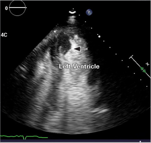
Initial apical 2-chamber transthoracic echocardiogram with an ultrasound-enhancing agent shows a thrombus measuring 2.7 × 1.3 cm (arrow) within the apex of the left ventricle. Not visualized is an additional 1.25 × 0.85 cm thrombus and the pseudoaneurysm measuring 8.7 × 7.6 cm.
Supplemental motion image is available for Figure 1.