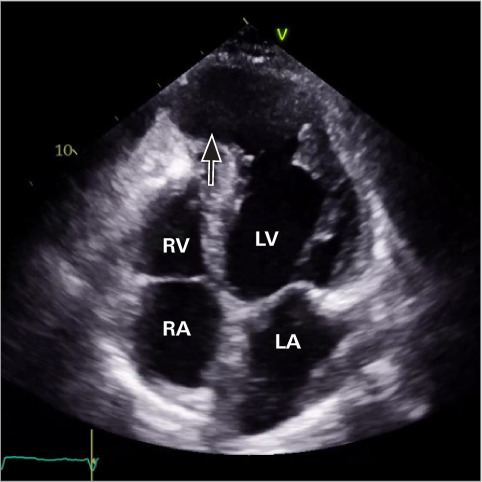
Fig. 3.

Repeat apical 4-chamber transthoracic echocardio-gram without UEA shows a large apical pseudoaneurysm (arrow) that measured 10.6 × 9.2 cm, 5 months after the index admission. The 2 prior thrombi had resolved but were also not visualized using UEA (thus not shown).
Supplemental motion image is available for Figure 3.
LA, left atrium; LV, left ventricle; RA, right atrium; RV, right ventricle; UEA, ultrasound-enhancing agent.
