Figure 6.
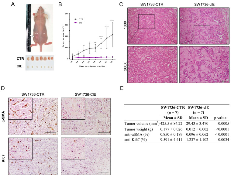
CRISPR/Cas9-mediated EZH2 gene editing inhibits ATC tumor growth in vivo. (A) Representative image of an animal injected with SW1736-CTR cells (in the left flank) and SW1736-ClE cells (in the right flank), and alignment of tumors collected from seven animals (n = 7) after euthanasia and surgical excision (each animal in a column). (B) Tumor volume evolution from day 18 (first visible tumor) to the euthanasia endpoint at day 35. (C) Histology of SW1736-CTR tumor (left) and SW1736-ClE tumor (right) stained with H&E. Magnification: 100× and 200×. (D) Immunohistochemical analysis of anti-αSMA (upper panel) and anti-Ki67 (lower panel) from section of SW1736-CTR (left) and SW1736-ClE (right) tumors counterstained with Gill’s hematoxylin at 200× magnification. Scale bar: 60 um. (E) Quantification of tumor parameters of volume and weight at the endpoint, and quantification of IHC staining for Ki67 and αSMA: Total and positive cells were counted for anti-Ki67 immunostaining and relative area for positive anti-αSMA area. *, p < 0.05; **, p < 0.01; ****, p < 0.0001 vs. SW1736-CTR.

