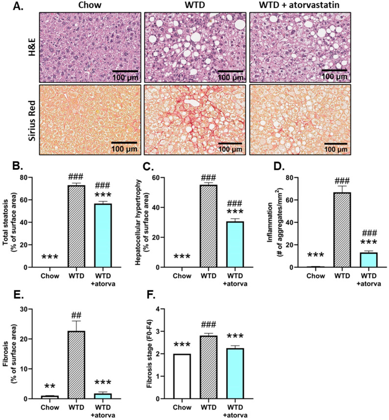
Figure 1.
Histological photomicrographs and quantitative analysis of NASH parameters. APOE*3-Leiden mice were fed a Western-type diet (WTD) with (n = 16) or without (n = 16) atorvastatin admix for 32 weeks. Chow-fed mice (n = 10) were included as a healthy reference. Representative histological photomicrographs of H&E-stained and Sirius Red-stained liver cross-sections (A). Total steatosis (B) and hepatocellular hypertrophy (C) as percentage of surface area, number of inflammatory foci per mm2 microscopic field (D), fibrosis as percentage of surface area (E) and fibrosis stage (F0–F4) (F) were determined at the study endpoint (t = 32 weeks) and are presented as mean ± SEM. ** p < 0.01, *** p < 0.001 vs. WTD. ## p < 0.01, ### p < 0.001 vs. chow.

