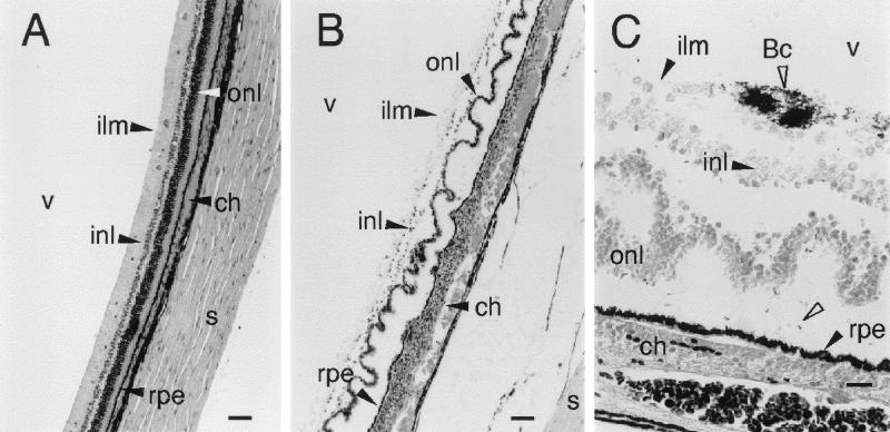
FIG. 4.
Retinal histopathology of experimental B. cereus endophthalmitis. (A) Control retina from eye injected with 0.1 ml of sterile saline. Stained with hematoxylin-eosin; bar, 100 μm. (B) Retinal and choroidal changes produced during B. cereus endophthalmitis. The outer nuclear layer shows extensive folding, and the choroid (ch) is engorged and heavily infiltrated with PMNs. PMNs can be seen crossing the retinal pigment epithelium (rpe) at local sites. Stained with hematoxylin-eosin; bar, 100 μm. (C) Magnified view of infected retina showing B. cereus (Bc; open arrowheads) aggregation at the surface of the retina and near the retinal pigment epithelium. Gram stain; bar, 20 μm. Abbreviations are defined in the legend to Fig. 3.

