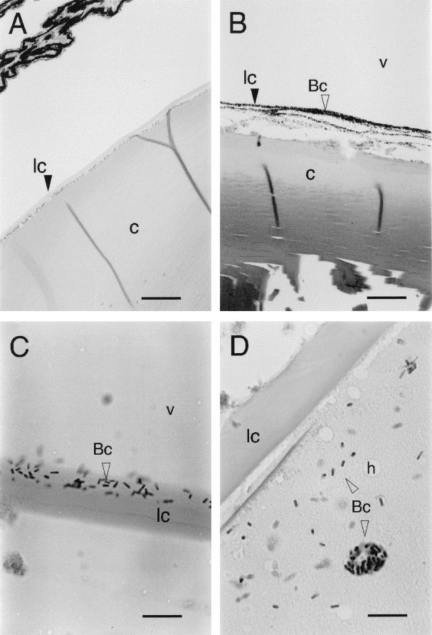
FIG. 5.
Progressive stages of B. cereus infiltration of rabbit lens during experimental endophthalmitis. (A) Control lens from eye injected with 0.1 ml of sterile saline. Bar, 200 μm. (B) Accumulation of B. cereus (Bc; open arrowhead) along the posterior margin of the lens 12 h postinfection. Bar, 100 μm. (C) B. cereus partially penetrating the posterior lens capsule. Bar, 20 μm. (D) Complete infiltration of lens cortex by B. cereus. Bar, 20 μm. Abbreviations: lc, lens capsule; c, lens cortex; v, vitreous fluid; h, hole. Open arrowheads point to B. cereus cells. All panels were stained with hematoxylin-eosin.

