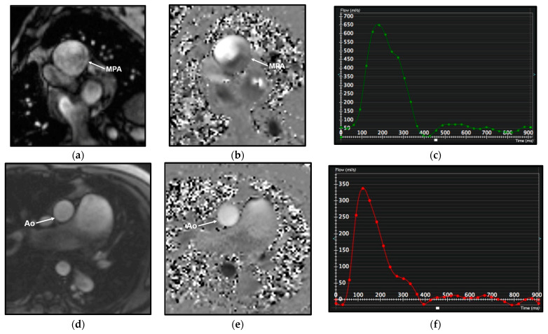
Figure 3.
Cardiac magnetic resonance (CMR) phase contrast imaging in the same patient as in Figure 2. Magnitude (a,d) and velocity (b,e) images in phase contrast imaging through the main pulmonary artery (MPA) and aorta (Ao) for flow quantification; note the increase of the main pulmonary artery caliber, larger than the aortic one. Graphical representation of flow velocity in the MPA (c) and in the Ao (f) (x-axis: time in msec; y-axis: flow velocity in mL/s) demonstrates a markedly increased flow in the MPA compared to the Ao, with a calculated pulmonary blood flow (Qp) of 9.96 L/min and a systemic blood flow (Qs) of 3.45 L/min, with a Qp/Qs ratio of 2.8, indicative of left-to-right shunt.

