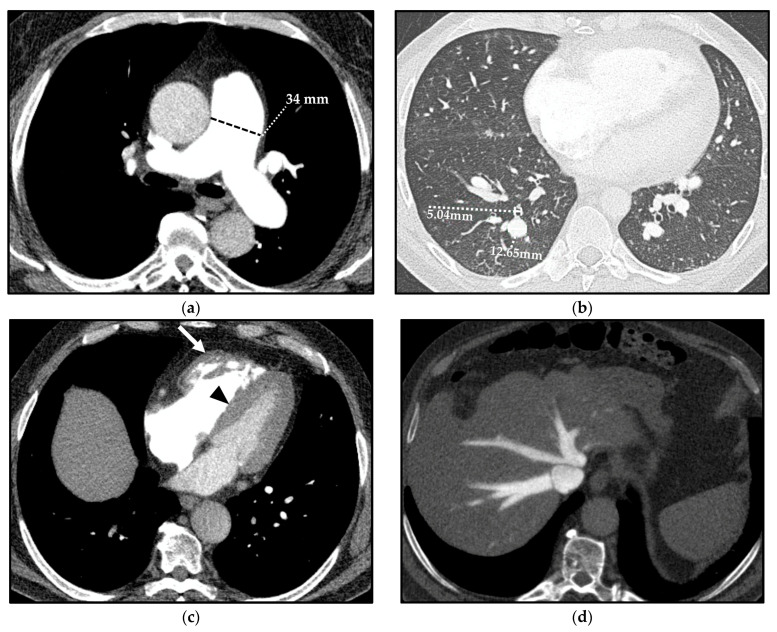
Figure 4.
Computed tomography (CT) indirect signs of pulmonary hypertension (PH). Vascular CT signs: main pulmonary artery (MPA) dilation (34 mm) (a) and increased segmental artery-to-bronchus ratio (>1) (b). Cardiac CT signs: right ventricle (RV) hypertrophy, with free wall thickness > 4 mm and trabecular hypertrophy (arrow), and flattening of the ventricular septum (arrowhead) (c). Extensive reflux of contrast medium into the inferior vena cava and hepatic veins (d).

