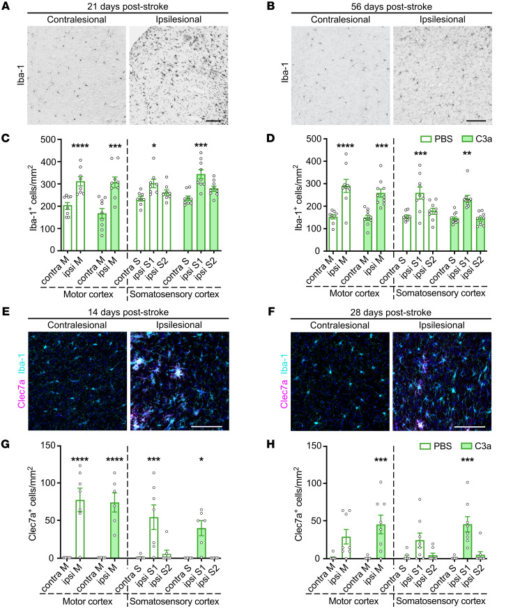
Figure 4. Intranasal C3a does not affect the density of microglia in peri-infarct cortex.
(A and B) Representative images of contralesional and ipsilesional cortex stained with antibodies against Iba-1 on P21 (A) and P56 (B). Cortical regions were chosen for analysis as shown in Figure 3D. Scale bars: 100 μm. (C and D) Density of Iba-1–positive cells in the proximal peri-infarct and contralesional cortex of mice treated with PBS or C3a on P21 (C) or P56 (D). PBS, n = 8–9; C3a, n = 9–10. (E and F) Representative images of contralesional and ipsilesional motor cortex stained with antibodies against Iba-1 and Clec7a on P14 (E) and P28 (F). Cortical regions were chosen for analysis as shown in Figure 3D. Scale bars: 100 μm. (G and H) Density of Clec7a-positive cells in the proximal peri-infarct and contralesional cortex of mice treated with PBS or C3a on P14 (G) or P28 (H). P14: PBS, n = 6; C3a, n = 6. P28: PBS, n = 10; C3a, n = 10. Bar plots represent mean ± SEM. Contra, contralesional; ipsi, ipsilesional; M, motor cortex; S, somatosensory cortex. Two-way ANOVA with Šidák’s planned comparisons: *P < 0.05, **P < 0.01, ***P < 0.001, ****P < 0.0001 for ipsilesional vs. contralesional comparisons.

