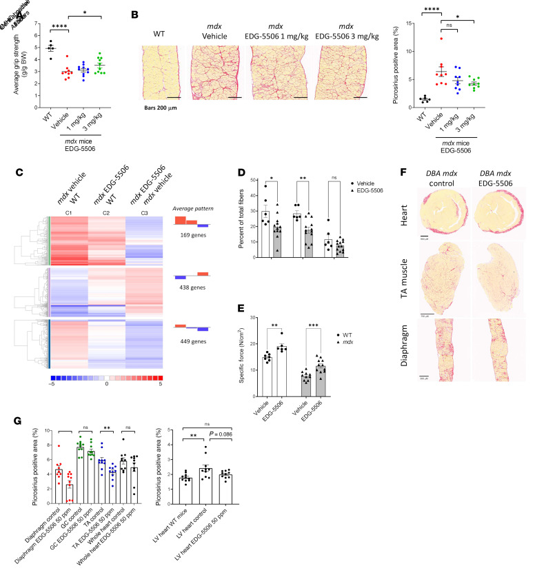
Figure 5. Longer-term exposure of protective levels of myosin inhibition are sufficient to decrease muscle degeneration and fibrosis in mdx mice.
(A) Average grip strength (experimenter blinded) measured after 5 weeks of dosing in mdx mice (n = 5–10). (B) Left: Representative images. Right: Quantification of collagen (stained with picrosirius red) in mdx mouse diaphragm after 8 weeks of treatment (n = 9–10). Scale bar: 200 μm. (C) RNA-Seq meta-analysis. Colors are graded by log2 fold change (WT, n = 2; mdx vehicle, EDG-5506, n = 3). (D) Histological quantification of central nuclei and eMHC-positive fibers in soleus muscle sections from post-weaning mdx mice after 3 weeks EDG-5506 administration. (E) Specific force in the soleus muscle ex vivo in postweaning mdx and WT mice after 3 weeks of treatment with EDG-5506 or vehicle. (F) Representative histology sections examining muscle fibrosis in DBA/2 mdx mice after 12 weeks of treatment with control or EDG-5506 chow (50 ppm or 0.13 mmol/Kg chow). Scale bar: 900 μm (heart); 700 μm (anterior tibialis [TA] muscle); 300 μm (diaphragm). (G) Quantification of collagen (picrosirius red area). Left: Collagen quantification in select muscles (GC, gastrocnemius). Right: Collagen quantification in the left ventricle (LV; n = 9–10). Data are shown as the mean ± SEM. Significance was calculated by 1-way ANOVA with Dunnett’s multiple-comparison test. *P < 0.05; **P < 0.01; ***P < 0.001; ****P < 0.0001.

