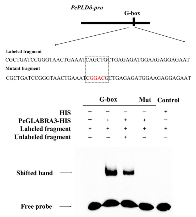
Figure 6.
Electrophoretic mobility shift assay (EMSA) verified the interaction of PeGLABRA3 with the PePLDδ promoter region. PeGLABRA3-HIS protein purified from prokaryotic expression was used for in vitro EMSA, while HIS-tagged protein was used as a negative control. The mutant probes (Mut, CAGCTG to CGGACG) were used to confirm the binding specificity of G-box to PeGLABRA3. The bases marked in red indicate the mutated bases in the mutant probe. In each loading panel, “+” and “−” indicate the presence or absence of protein and probe, respectively. The cold probe concentration was 10× and the concentration of the polyacrylamide gel was 6%. The EMSA experiment was repeated three times and the representative images are shown.

