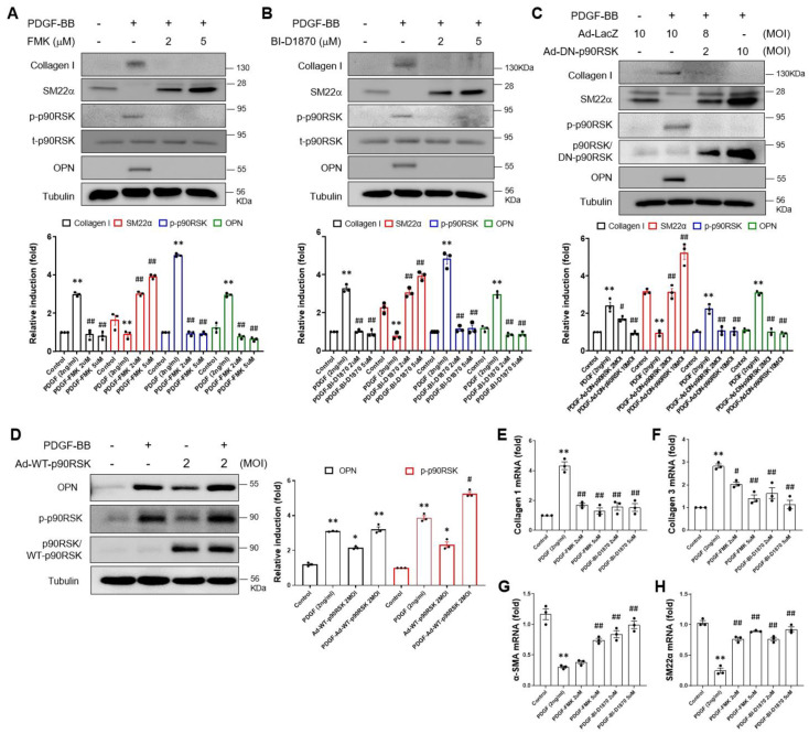
Figure 2.
p90RSK is involved in PDGF-BB-mediated phenotypic switching of VSMCs. (A,B) VSMCs were treated for 1 h with FMK and BI-D1870, followed by treatment with 2 ng/mL of PDGF-BB for 24 h. The expression level of collagen I, SM22α, p90RSK, and OPN proteins was determined via immunoblotting. The results of the densitometric analysis of immunoblotting bands were presented in the bar graphs. ** p < 0.01 vs. vehicle control (lane 1), ## p < 0.01 vs. cells treated with PDGF-BB (lane 2). (C,D) VSMCs were transduced either with Ad-WT-RSK and Ad-DN-RSK or Ad-LacZ as a control for 24 h, followed by treatment with 2 ng/mL of PDGF-BB for 24 h. The level of protein expression was analyzed via immunoblotting. * p < 0.05, ** p < 0.01 vs. vehicle control (lane 1), # p < 0.05, ## p < 0.01 vs. cells treated with PDGF-BB (lane 2). (E–H) The mRNA expression level of collagen I, collagen III, α-SMA, and SM22α was determined by quantitative RT-PCR assay in VSMCs treated with FMK or BI-D1870. VSMC was pretreated with either 2 or 5 μM of FMK or BI-D1870 for 1 h, followed by treatment with 2 ng/mL of PDGF-BB for 24 h. The data are representative of the three independent experiments. ** p < 0.01 vs. vehicle control (lane 1), # p < 0.05, ## p < 0.01 vs. cells treated with PDGF-BB (lane 2).

