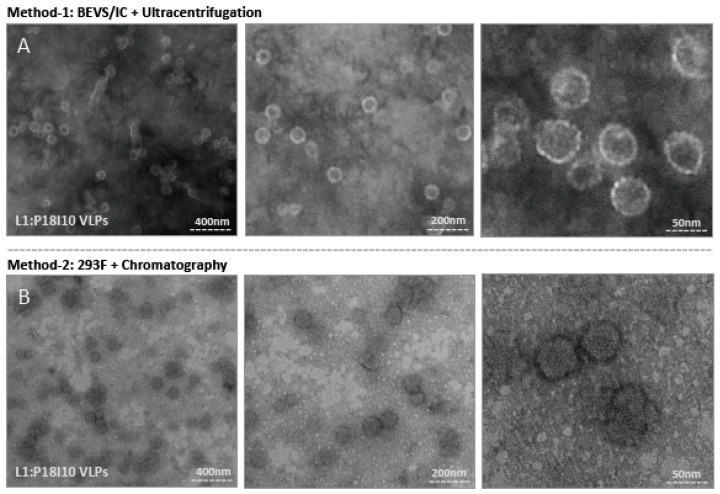
Figure 5.
Electron micrographs of purified L1:P18I10 VLPs. (A) Morphology of ultracentrifugation-purified L1:P18I10 VLPs. L1:P18I10 VLPs were equilibrated with PBS, absorbed on UV-charged carbon-coated copper grids, and negatively stained with 2% PTA. (B) Morphology of chromatography-purified L1:P18I10 VLPs. L1:P18I10 VLPs were equilibrated with Tris-HCl, absorbed on UV-charged carbon-coated copper grids, and negatively stained with 2% uranyl acetate. Images were acquired under transmission electron microscopy Tecnai Spirit 120 kV. The bar represents 50 nm at magnification SA270K (left panel), 200 nm at magnification SA59000 (middle panel) and 400 nm at magnification SA529500 (right panel).

