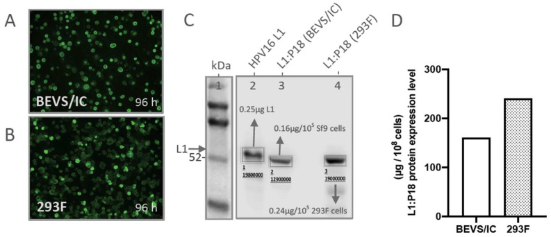
Figure A1.
L1:P18I10 proteins production and transfection efficiency by using BEVS/IC or 293F expression systems. (A,B) Immunofluorescence staining of L1:P18I10 proteins produced from BEVS/IC and 293F systems. Sf9 cells (top panel) and 293F cells (bottom panel) were harvested in day 4 post-transfection. Cells were probed with anti-HPV16 L1 mAb and detected with anti-mouse IgG-FITC (green channel). (C) Quantification Western blot analysis of L1:P18I10 proteins produced from BEVS/IC and 293F systems. A total of 1 × 10⁵ Sf9 or 293F cells in day 4 post-transfection were collected and analyzed by Western blot stained with anti-HPV16 L1 mAb. The expression level of L1:P18I10 proteins was densitometrically quantified by Image Studio Lite 5x software. The HPV16 L1 proteins were used as a control for quantification. Lane 1: protein molecular weight marker; Lane 2: 0.25 µg HPV16 L1 protein; Lane 3: BEVS/IC-produced L1:P18 protein; Lane 4: 293F-produced L1:P18 protein. (D) Comparison of L1:P18I10 protein expression level between BEVS/IC and 293F expression systems. The expression level of L1:P18I10 proteins was densitometrically quantified by Image Studio Lite 5.x software. The HPV16 L1 proteins were used as a control for quantification.

