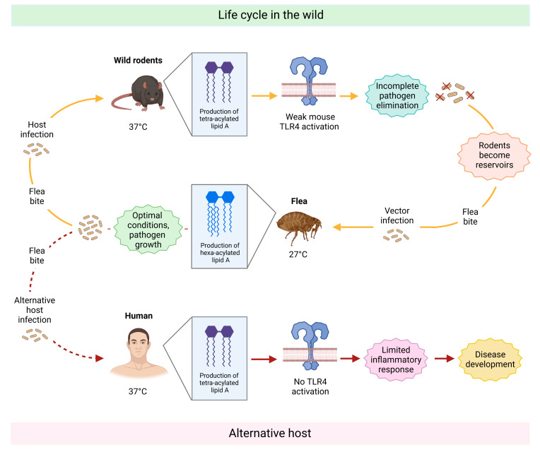
Figure 4.
Schematic representation of Yersinia pestis life cycle with focus on how the lipid A moiety changes during the different stages, ultimately interfering in the host’s immune response. The top part of the image shows the life cycle in wild animals. Briefly, Yersinia pestis-infected fleas bite wild rodents and transmit the pathogen. Triggered by the rodent’s body temperature of 37 °C, it starts producing a less immunogenic variant of lipid A (tetra-acylated), which is not able to elicit an effective immune response. As a result, Yersinia pestis is not fully eliminated and persists. The rodent becomes a reservoir of the pathogen in the wild and can, in turn, infect vector fleas, which feed on their blood. Once Yersinia pestis reaches the flea digestive tract (27 °C), it produces a hexa-acylated form of lipid A and starts proliferation in these favored conditions. By biting a new host, the flea can further spread the disease. Once humans are accidentally bitten (bottom part of the figure), the bacterium produces the tetra-acylated form of lipid A again to adapt to the human body temperature. However, in contrast to rodent TLR4, the human TLR4 does not recognize this lipid A variant, and therefore, it is not able to activate the downstream signaling cascade. As a result, the immune response is ineffective as it has to rely only on other defense mechanisms. Yersinia pestis invades the body undisturbed, causing a disease called plague.

