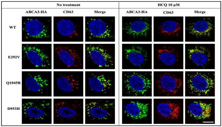
Figure 5.
A549 cells stably expressing WT or mutated ABCA3-HA were treated with RPMI-1640 + 10% FBS (no treatment) or RPMI-1640 + 10% FBS-added HCQ 10 µM (HCQ 10 µM) for 24 h, and then stained for ABCA3-HA and lysosomal marker CD63. ABCA3-HA in green, CD63 in red, and DAPI in blue. Scale bar represents 20 μm.

